Mechanical ventilation and good critical care are mainstays of therapy when treating patients with ARDS.
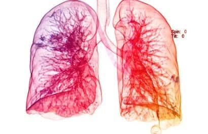
In the nearly 3 decades since the initial description of the adult respiratory distress syndrome (ARDS), with its attendant mortality of 50% to 70% (and 90% mortality for patients with concomitant sepsis),1 substantial advances in the understanding of the disease process have occurred. Despite intensive investigations centered on the pathophysiology and potential pharmacotherapy of ARDS, no wonder drug exists. In spite of current critical care practice, little improvement has been made in overall mortality. Management of patients with ARDS remains primarily supportive, with mechanical ventilation and good critical care the mainstays of therapy. As new treatment modalities are evaluated, however, subsets of ARDS patients showing improvement in morbidity and mortality continue to be identified. Investigation of neonatal and pediatric respiratory distress has also yielded new therapeutic strategies that may affect the adult population (Figure 1, page 48). Various treatment strategies include pressure-limited ventilation (permissive hypercapnia), inverse-ratio ventilation (IRV), high-frequency jet ventilation (HFJV), high-frequency oscillatory ventilation (HFOV), intratracheal pulmonary ventilation, and prone positioning. Cutting-edge therapies, such as partial liquid ventilation and inhaled nitric oxide, provide additional treatment modalities. A component of some (or all) of these strategies will find a role in clinical practice.
Definition
In 1967, Ashbaugh et al1 described a series of 12 patients suffering from acute-onset tachypnea, hypoxemia, and reduced pulmonary compliance; patchy, diffuse infiltrates were visible in their chest radiographs. This syndrome was named ARDS. In the three intervening decades, many additional features have been noted, yet there is still no uniformly accepted definition. Originally, the clinical criteria included hypoxia, decreased pulmonary compliance, and an abnormal chest radiograph. Routine right-heart hemodynamic monitoring led to the inclusion of normal left ventricular filling pressure to distinguish congestive heart failure from ARDS. In 1988, Murray et al2 proposed their lung-injury score (Figure 2, page 50) in an attempt to standardize the assessment of what was quickly becoming a leading cause of mortality in intensive care units (ICUs). The lung-injury score added the degree of positive end-expiratory pressure (PEEP) to the original three criteria and devised a scoring system associated with varying degrees of severity within each parameter. A modified lung-injury score and a proposal by the American-European Consensus Conference have since added to the controversy (Figure 3, page 51).3
Many clinical trials have relied on differing criteria to define respiratory failure and/or ARDS, making it difficult to draw direct conclusions about the treatments proposed. A recent prospective, randomized comparative study4 analyzed the accuracy of the lung-injury score, the modified lung-injury score, and the American-European Consensus Conference definition in comparison with the original definition of ARDS. All three were found to identify similar patients, provided their criteria were applied to patients with clearly defined at-risk diagnoses for ARDS. Currently, ARDS is most frequently defined through the identification of an inciting event (or events) in a patient presumed to be at risk, in addition to the following clinical criteria: severe compromise in systemic oxygenation with Pao2/Fio2 ratio < 200, diffuse bilateral infiltrates (whiteout) seen in chest radiographs, and severe reduction of pulmonary compliance.
Epidemiology
The annual incidence of ARDS, as identified by the American-European Consensus Conference,3 is 5 to 10 per 100,000. This number is not, however, spread evenly within the predisposed groups. For example, Messent et al5 reported that only 1.5% of patients undergoing cardiopulmonary bypass developed ARDS, while Sloane et al6 reported a much higher incidence in patients with sepsis. Furthermore, patients with trauma-induced ARDS are more likely to survive than those with an infective etiology.7 Therefore, the precipitating condition defines both epidemiology and survivability.
Pathophysiology
ARDS is the manifestation of progressive pulmonary inflammation incited by a variety of pulmonary and extrapulmonary insults. Of particular importance are sepsis, trauma, and injury due to mechanical ventilation (barotrauma and volutrauma) associated with widespread activation of a systemic inflammatory response.8 This response leads to parenchymal lung injury, increased permeability of the alveolar-capillary barrier, and deactivation of surfactant.9 Damage to the pulmonary vasculature also occurs through the release of free radicals of oxygen, vasoactive peptides, and tissue proteases.
Bone10 noted that the list of proinflammatory mediators contributing to the systemic inflammatory response syndrome (SIRS) has expanded greatly in recent years. Among the first of these endogenous mediators to be identified were tumor necrosis factor and interleukin 1 (IL-1). The list has been expanded to include many other cytokines, the products of neutrophil degranulation, platelets and the clotting factors formed on their surfaces, complement fragments, platelet-activating factor, and arachidonic-acid derivatives. A new class of mediators, the chemokines (which exert potent chemotactic and activating effects on leukocytes), has been added to the list, and other factors undoubtedly await discovery. As these mediators have been studied, much has been learned about how circulating cells, such as macrophages, neutrophils, and platelets, are recruited to a site of injury or infection, and how these circulating cells are able to communicate with, and attach to, the endothelium. The endothelium also contributes to its own defense and repair, and intracellular junctions can expand or contract in response to a variety of stimuli.
Over the past several years, multiple clinical trials have examined different methods of counteracting proinflammatory mediators. All of these trials failed to demonstrate any survival benefit attributable to treatment. Many investigators interpret these trials as underestimating the magnitude, complexity, and ongoing nature of the inciting event for the inflammatory response. Evidence is accumulating that, in response to the original inciting event (the inflammatory response), the body also mounts an anti-inflammatory response. Agents identified so far as participating in this anti-inflammatory response include IL-4, IL-10, IL-11, and IL-13; transforming growth factor-b; colony stimulating factors; soluble receptors for tumor necrosis factors; and receptor antagonists to IL-1. If the compensatory anti-inflammatory response is sufficiently severe, it will be manifested clinically as anergy, an increased susceptibility to infection, or both. If balance cannot be established and homeostasis is not restored, a massive proinflammatory reaction (SIRS) or an anti-inflammatory reaction will ensue. A range of clinical sequelae may then follow, including ARDS.
Once an initializing event or mediator induces increased alveolar permeability, the progression of the lung injury is usually divided into three phases: exudative, proliferative, and fibrotic.11 The exudative phase generally lasts for up to a week and is closely tied to the inciting systemic events. It is during this phase that interstitial and alveolar edema leads to atelectasis, hyaline membrane formation, alveolar collapse, and reduction in overall lung compliance. This pathology is mainly located in the lower, dependent regions of the lung. The perfusion of this consolidated lung tissue, coupled with the redistribution of ventilation to the upper, nondependent regions of the lung, causes a large shunt and, thus, a severe gas-exchange impairment.12 The proliferative phase follows, with a gradual reduction in systemic inflammation and a resultant proliferation of type II pneumocytes in an attempt to take reparative measures. Edema and inflammatory cell infiltration persist and, in the later part of this phase (which may last for up to several weeks), fibrosis begins. The fibrotic phase is characterized by further deposition of collagen in both the interstitium and the air spaces of collapsed alveoli.
The time course of ARDS is variable, and the determinants of outcome are not well understood. The overall mortality of patients with advanced disease (a prominent proliferative infiltrate and severe fibrosis) ranges from 50% to 90%. Survivors with short time courses (2 to 5 days) have relatively limited exudative and proliferative phases, and the resultant fibrotic phase produces little to no long-term residual parenchymal scarring. Protracted cases are associated with more severe lung injury and often progress to extensive fibrosis and irreversible pulmonary dysfunction.
Mechanical Ventilation
The use of positive-pressure ventilation in severe respiratory failure has undergone a fundamental change in recent years, based on extensive human and animal studies that suggested that mechanical ventilation may be a major contributor to the perpetuation of ARDS. Regional pressure-related overdistention of healthy alveoli can result in a pattern of diffuse alveolar damage that is histologically indistinguishable from ARDS having other causes.13 Ranieri et al8 demonstrated elevated plasma and bronchoalveolar-lavage concentrations of inflammatory mediators after treatment with conventional mechanical ventilation. Increases in capillary permeability, edema, and inactivation of surfactant have also been demonstrated.14 Animal studies have suggested that alveolar overdistention, whether produced by positive or negative pressure, is responsible for the initiation and/or propagation of the injury. Transpulmonary pressures of approximately 35 cm H2O result in maximal alveolar volume in normal lungs, and the use of distending pressures in excess of this level risks parenchymal injury. Further, mechanical ventilation can prolong an otherwise limited ARDS injury and lead to a more fulminant and severe course.
Mechanical-ventilation management is now based on preventing overdistension of normal alveolar tissue when peak airway pressures reach levels higher than 35 to 40 cm H2O. This has led to the development of techniques to improve oxygenation via the use of adjunctive measures or through the adaptation of current ventilator strategies to include therapies that minimize the secondary injury known to occur.
Permissive Hypercapnia
The standard approach to mechanical ventilation has been to maintain normal Paco2 and Pao2 by applying tidal volumes of 10 to 15 mL/kg and minimizing Fio2. In order to reduce potential ventilator-induced injury, management approaches have veered away from blood-gas–oriented results toward points based on moderation in peak inspiratory pressure (PIP). When the reduction of PIP, rather than achieving a normal Pco2, becomes the primary goal, it can be achieved using either volume-control or pressure-control modes. The use of respiratory rates of less than 20 breaths per minute can further limit shear forces and the generation of autoPEEP. In severe ARDS, the level of alveolar ventilation achieved is insufficient to maintain a normal Pco2, and hypercapnia results. If pressure limitation is accepted as the primary goal, then carbon dioxide levels are permitted to rise, if necessary. A tidal volume of as little as 5 mL/kg is used in order to maintain PIP below a commonly accepted threshold for injury of 35 to 40 cm H2O.15 Clinical studies16 in humans have demonstrated that hypercapnia is well tolerated, and patients undergoing this strategy have improved survival, compared with predicted mortality.
Problems associated with permissive hypercapnia seem related to the physiologic response to an increasing carbon dioxide level and the resulting systemic respiratory acidosis.17 The majority of sequelae stem from an increase in sympathetic nervous system activity. Tachycardia, increased cardiac output, decreased peripheral resistance, and increased splanchnic blood flow are frequently associated with permissive hypercapnia. Increased cerebral blood flow is also well described, with the potential for increased intracranial pressure. Direct myocardial suppression by carbon dioxide is usually counterbalanced by stimulation of catecholamine release from the adrenal medulla. Often, management of an ARDS patient in the ICU setting requires sedation and/or neuromuscular paralysis to block the reflexive respiratory drive. In some research, buffering the acute acidosis has not been routine, and Hickling et al16 reported no adverse effects in patients with a pH as low as 7 and a Pco2 of 120 mm Hg. Use of sodium bicarbonate may be limited to selected patients in whom hypercapnia results in intolerable cardiovascular or cerebral compromises. Permissive hypercapnia is best tolerated when imposed gradually over a period of several hours. Severe hypercapnia can be efficiently reduced using a combination of respiratory washout, an increased respiratory rate, and reduced instrumental dead space.18 The potential benefits, physiologic characteristics, and relative safety lend support to permissive hypercapnia in patients not at risk for potential worsening of marginal hemodynamic status.16,19
Using IRV
Standard mechanical ventilation attempts to simulate normal physiology, in which inspiration is considerably shorter than expiration. The usual ratio of inspiration to expiration (I:E) is in the range of 1:2 to 1:3. During the inspiratory phase of mechanical ventilation, positive pressure is used to overcome the impedance of the breathing circuit and conducting airways, as well as to inflate the terminal alveoli. To prevent unstable alveoli with reduced compliance from collapsing during the expiratory phase, PEEP maintains persistent distending pressures in the airway, thereby promoting alveolar recruitment, increasing end-expiratory lung volume, and improving oxygenation. The combination of PIP, PEEP, I:E ratio, and gas-flow rate determines the mean airway pressure (MAP), which represents overall airway pressures averaged over time during the respiratory cycle.
Prolonging the inspiratory time has also been advocated for overcoming regional inhomogeneities in oxygenation to improve arterial oxygenation. Increasing inspiratory time to a point at which the I:E exceeds 1:1 is called IRV. The alveoli are maintained at a higher volume for a greater portion of the respiratory cycle. In addition, since the expiratory phase is shortened, air trapping occurs, providing autoPEEP. These phenomena cause recruitment of collapsed or consolidated lung tissue and a redistribution of inspired gas towards more dependent regions of the lung, thereby decreasing the intrapulmonary shunt.12 In addition, the reduction in the repetitive opening and closing of unstable lung units causes a reduction in the shear forces applied to the parenchyma, thereby minimizing ventilator-induced lung injury.
IRV has potential problems, however. Whereas some investigators feel that autoPEEP is a necessary component in recruiting unstable alveoli, others propose that it results in regional overdistention, with resultant barotrauma and possible pneumothorax. Furthermore, the increase in mean airway pressure can result in marked elevations in intrathoracic pressure, impaired venous return, and reduced cardiac output, with resultant hemodynamic compromise in a patient who is already critically ill.
Most experience with IRV in the treatment of acute respiratory failure is limited to the pressure-control mode (PC-IRV).20 The majority of studies show that PIP can be reduced without a reduction in alveolar ventilation; however, no improvement in mortality was demonstrated by switching to PC-IRV.21 Unfortunately, the majority of patients were initially supported using a volume-controlled ventilator algorithm with high PIP and Fio2 for prolonged periods prior to the change to PC-IRV. In order to demonstrate the relative merits of PC-IRV, it will be necessary to conduct a prospective, randomized trial in patients with similar severities of illness who are enrolled early enough in the course of treatment to reveal any potential improvement in survival. At this time, the use of PC-IRV as an alternative to traditional volume-controlled mechanical ventilation remains unproven as a method to improve the morbidity/mortality of respiratory failure.
High-Frequency Ventilation
High-frequency ventilation (HFV) involves the delivery of respiratory tidal volumes averaging significantly less than anatomic dead space at a high frequency. Typical tidal volumes are in the range of 1 to 3 mL/kg with a respiratory rate of 150 to 300 breaths per minute. When compared with conventional ventilation, HFV improves oxygenation by increasing mean airway pressure and decreasing PIP due to the reduction in tidal volumes; theoretically, a reduction in iatrogenic barotrauma is expected.22 The maintenance of adequate mean airway pressures for adequate gas exchange without elevated peak pressures has been shown to be beneficial in infants with respiratory distress syndrome (RDS).23
HFOV uses a reciprocating piston, diaphragm, or bellows to generate a sinusoidal respiratory waveform during the breathing cycle. Characteristically, both the inspiratory and expiratory phases are active, with gas driven into and withdrawn from the lungs by the pump stroke. The frequency of oscillation ranges over a wide spectrum (1 to 50 cycles per second), and pumps with variable I:E ratios are available. Results of several randomized studies using HFOV have demonstrated a decreased incidence of barotrauma and bronchopulmonary dysplasia, as well as a reduction in the number of term infants who deteriorate to a point at which they require extracorporeal membrane oxygenation (ECMO).24 The use of HFOV allows improved control of Pco2, especially for the management of persistent pulmonary hypertension in the neonate (where intentional respiratory alkalosis can reverse a right-to-left ductal shunt or persistent fetal circulation). The improvements in oxygenation can be attributed to static inflation (up to 30 cm H2O) with intermittent increases to assist in alveolar recruitment and re-expansion of atelectatic segments. The most conclusive study,25 however, demonstrated that HFOV resulted in a significantly higher incidence of intraventricular hemorrhage in neonates (and in other long-term sequelae) without providing a clear benefit over controls. This mode of therapy has mainly been used as a last alternative prior to the initiation of ECMO, or as a rescue technique when infants have been failed by either conventional ventilation or an alternative advanced technique. Isolated spectacular responses in individual patients continue to stimulate further investigation of this therapy.
HFJV consists of the intermittent delivery of gas through a small-bore cannula positioned within the airway that delivers fresh gas in short bursts. A cycling mechanism is employed that allows regulation of rate, inspiratory time, and driving pressure. Rates are commonly 100 to 200 breaths per minute, and the expiratory phase of the cycle is entirely passive, depending on chest-wall and lung compliance. The primary use of HFJV is as a rescue therapy in adults with hypoxemia related to acute respiratory failure in the face of high PEEP or a significant air leak. The use of smaller tidal volumes with increases in mean airway pressure, but not PIP, is purported to be helpful in avoidance of worsening barotrauma. Carlo et al26 reported on a group of neonates with severe RDS and showed that HFJV produced a significant reduction in mean airway pressure and Pco2, but no variation in mortality, the incidence of air leaks, or the duration of assisted ventilation, when compared with conventional ventilation. Complications and risks associated with HFJV include tracheal obstruction and inflammatory tracheal injuries. Successful application has been achieved in neonates with RDS due to meconium aspiration or congenital diaphragmatic hernias.27 Most responders to HFJV show the greatest improvement within the first few hours of treatment.
Intratracheal Pulmonary Ventilation
A unique recent innovation intended to reduce the volutrauma/barotrauma of volume ventilation was developed by Muller et al28 using a specially designed endotracheal tube with a conventional ventilator. The endotracheal tube operates with continuous oxygen flow delivered at the level of the carina. The design is such that a Venturi effect produces efficient carbon dioxide clearance with a tidal volume that is a fraction of conventional volumes; very low airway pressure is maintained. The ventilator itself is used only for the expiratory phase, while the catheter delivers fresh gas. The system has been extensively studied in animals,29 and there have been human case reports.30 Measurements of end-expiratory pressures demonstrated no inadvertent PEEP. Although promising, this method has yet to be evaluated in large, randomized outcomes studies.
Prone Positioning
When a patient is mechanically ventilated in the supine position, there is preferential perfusion of the dependent lung regions due to the effects of gravity, as well as the impact of the mechanical distention of capillaries in the dependent (or basal) portions of the pulmonary parenchyma. Several studies,31-35 in animals and humans, have demonstrated a significant improvement in oxygenation and ventilation-perfusion inequality when ventilation is carried out in the prone position. Mechanisms for this improvement are an increase in functional residual capacity, a change in diaphragm motion, and a redistribution of blood flow to less-affected lung regions (resulting in increased recruitment of previously atelectatic, but uninjured, lung units). The gravitational distribution of pleural pressure is more uniform in the prone position. Jolliet et al33 demonstrated a rapid improvement in responders (57%), with the beneficial effect not disappearing immediately on return to the supine position. They also demonstrated no deleterious effects on hypoxemia or hemodynamics in nonresponders. Ventilating a patient in the prone position presents unique nursing and resuscitative challenges, however, and may not always be well tolerated by a patient with marginal hemodynamic status.
Surfactant Replacement
Surfactant is known to decrease alveolar surface tension and alveolar edema and to exhibit some anticytokine effects. Surfactant is inactivated during ARDS, probably due to proteolysis from neutrophil elastase. ARDS is also associated with a massive influx of activated neutrophils. Baker et al9 demonstrated a similar pattern of in vivo cleavage for surfactant-specific protein A and in vitro neutrophil elastase cleavage for surfactant-specific proteins. The safety of aerosolized surfactant has been demonstrated,36 but the results of a number of trials are mixed. Recent studies indicate that surfactant therapy provides significant improvements in oxygenation with much less lung injury than conventional ventilation in ARDS patients.37 Marraro et al38 found the efficacy of surfactant therapy to be strictly connected with early treatment. In contrast, Anzueto et al39 investigated surfactant use in sepsis-related adult ARDS patients and reported no improvement in 30-day survival, length of ICU stay, duration of mechanical ventilation, or physiological function. Controversies remain regarding routes of administration, the use of natural versus artificial or recombinant surfactants, and dosages. Combining surfactant therapy with antiprotease therapy may improve therapeutic prospects.9
Partial Liquid Ventilation
Perfluorocarbons (PFCs) have unique gas-solubility properties, combined with low vapor pressure, relatively high density, and low surface tension. In addition, they are biologically inactive, are physiologically inert, are insoluble in lipids and water, and have limited potential for systemic absorption. PFCs have several proposed modes of action that may affect ARDS; however, their significance has yet to be rigorously determined. When instilled into the airway, PFCs are distributed primarily to the dependent regions of the lung by virtue of their density (which is greater than that of water). They displace the inflammatory exudates, acting as a lavage, while possibly improving lung compliance via surfactant-like properties. In a rabbit model of acute lung injury, Rotta et al40 demonstrated a significant decrease in lung-injury scores in a group ventilated using partial liquid ventilation with perfluorooctyl bromide, compared with a group ventilated using controlled minute ventilation. The injury scores for the two groups were similar in the nondependent lung regions, but were significantly different in the dependent lung regions. PFCs may help to redistribute pulmonary perfusion from the dependent, collapsed alveoli to better-ventilated, nondependent regions. PFCs are bacteriostatic and may also have anti-inflammatory activity. In animal models of ARDS, researchers have demonstrated the efficacy of PFCs for improving gas exchange and pulmonary function,41 with reductions in physiologic shunting and increases in compliance.
Human studies have been initiated in pediatric patient populations, with PFC instillation to the predicted functional residual capacity of 30 mL/kg in combination with standard gas ventilation. Partial liquid ventilation was able to support gas exchange and allow some improvement in lung compliance.42 A subsequent trial in infants with acute respiratory failure in whom conventional therapy had failed showed that all patients demonstrated some improvement in oxygenation without serious adverse events.43 A multicenter phase I and II trial of partial liquid ventilation in adults with ARDS demonstrated the safety, and the few related severe adverse effects, of partial liquid ventilation. Improvement of gas exchange was observed in this series of patients over the 48 hours after initiation of partial liquid ventilation.44 Effects on gas exchange and outcomes, however, have been inconsistent. Again, isolated spectacular responses in individual patients have been reported. Partial liquid ventilation may be a viable alternative for infants with RDS, but its role in ARDS awaits the results of further investigations.
Inhaled Nitric Oxide
ARDS is characterized by progressive hypoxemia secondary to intrapulmonary shunting. Reflex pulmonary vasoconstriction in response to this hypoxemia increases intrapulmonary resistance, induces microvascular thrombosis in the regions of stagnant flow, and potentially overloads the right heart in a critically ill patient. Reducing pulmonary arterial resistance should enhance both flow and gas exchange efficiency, and this reduction has been the focus of a number of animal and clinical studies. Early work with intravenous pulmonary vasodilators was largely unsatisfying because of the unwanted side effect of systemic hypotension. This, combined with the resultant increase in blood flow to regions in the lung that were either collapsed or filled with fluid, increased ventilation-perfusion mismatching and ultimately worsened hypoxia.
In the late 1980s, nitric oxide was reported to be a potent, pulmonary-selective vasodilator. The vascular endothelium plays a vital role in the generation of vascular relaxing factors and is responsible for regulation of both arterial and venous smooth muscle tone. When administered via inhalation, nitric oxide is distributed to regions of the lung that contain patent alveoli and are able to transfer nitric oxide to the bloodstream. Through diffusion to the surrounding vasculature, blood flow is increased to those areas of the parenchyma capable of gas exchange, and blood is diverted away from poorly ventilated areas. These effects are manifested as an increase in arterial oxygenation and a reduction in ventilation-perfusion mismatching. Nitric oxide has an affinity for hemoglobin that is on the order of 1,500 times that of carbon monoxide. Upon combining with hemoglobin, nitric oxide is immediately inactivated and, therefore, systemic effects are rarely seen.
Potential toxicities of nitric oxide therapy are related to the formation of nitrogen dioxide and methemoglobin. Approximately 80% to 90% of inhaled nitric oxide is absorbed within the bloodstream, and it combines to form methemoglobin. The factors affecting the rate of conversion are related primarily to the dose of nitric oxide, hemoglobin level, and oxygen saturation. Doses of up to 80 ppm have been investigated, and minimal toxicity from methemoglobinemia has been shown. Under conditions of antioxidant depletion, nitric oxide can induce radical formation and subsequent inflammation.
Nitric oxide is used as rescue therapy in newborns with severe hypoxia secondary to severe respiratory failure in an attempt to prevent the need for ECMO.45 Nitric oxide has also been used to treat the transient, severe pulmonary hypertension seen following lung transplantation. Currently, inhaled nitric oxide is part of ongoing studies as an adjunct in the treatment of adult ARDS. Response rates (defined as increases of 20% to 25% in Pao2) of 36% to 42% in supine patients have been reported.34,46 Martinez et al34 reported a higher response rate (57%) to nitric oxide therapy as an additive effect in prone patients. Furthermore, Iotti et al46 demonstrated that a nitric-oxide dose of 5 ppm was optimal for obtaining the plateau increase in arterial oxygen content. The Pao2 was shown to be higher in some patients at higher doses, but the oxygen content was not increased (due to an increase in methemoglobin at higher concentrations of nitric oxide).46 Despite transient reductions in peak airway pressures and inspired oxygen levels, however, nitric oxide therapy may or may not have an effect on overall mortality in an ARDS population with frequent concomitant illnesses and/or injuries, each with its own attendant morbidity. The preliminary results of a randomized, controlled, blinded trial in non-septic ARDS patients (using nitric oxide doses of 0, 1.25, 5, 20, 40, and 80 ppm) have shown no differences in outcomes.47
Conclusion
At present, the body of knowledge related to the management of the adult, child, or neonate with respiratory failure is rapidly expanding. With the introduction of novel treatment approaches, the revisitation of previously used approaches, and improvements in understanding of the pathophysiology of ARDS, investigators strive to develop the ideal treatment with a minimum of ill effects. A number of therapies appear to be promising, pending controlled studies, while others fade as no benefits are shown. Several concepts have been persistent: ARDS is multifactorial and complex, and may be the end-organ response to a host of initiating events; pressure-controlled or gentle ventilation limits barotrauma/volutrauma during the management of ARDS; several modalities (surfactant, nitric oxide, and partial liquid ventilation) appear to affect outcomes in selected patients; and, given the complexity of the problem, each case of RDS is still unique, and treatment should remain individualized. All new treatment modalities, however, must be subjected to prospective, randomized trials. The clinician must be knowledgeable about all of these treatment options, as individual patients may show remarkable responses to one or more of them.
Joseph B. Zwischenberger, MD, is professor of surgery, medicine, and radiology and director of the general thoracic surgery and extracorporeal membrane oxygenation programs, Division of Cardiothoracic Surgery, University of Texas Medical Branch, Galveston. Scott K. Alpard, MD, is a surgical research fellow in the division. Akhil Bidani, MD, PhD, is professor and chief of the Division of Pulmonary and Critical Care Medicine at the university. Pablo Pritchard is a medical student at the university.
References
1. Ashbaugh DG, Bigelow DB, Petty TL, Levine BE. Acute respiratory distress in adults. Lancet. 1967;II:319-323.
2. Murray JF, Matthay MA, Luce JM, Flick MR. An expanded definition of the adult respiratory distress syndrome. Am Rev Respir Dis. 1988;138:720-723.
3. Bernard GR, Artigas A, Brigham KL, et al. The American-European Consensus Conference on ARDS. Definitions, mechanisms, relevant outcomes, and clinical trial coordination. Am J Respir Crit Care Med. 1994;149:818-824.
4. Moss M, Goodman PL, Heinig M, Barkin S, Ackerson L, Parsons PE. Establishing the relative accuracy of those definitions of the adult respiratory distress syndrome. Crit Care Med. 1995;23:1629-1637.
5. Messent M, Sullivan K, Keogh BF, Morgan CJ, Evans TW. Adult respiratory distress syndrome following cardiopulmonary bypass: incidence and prediction. Anaesthesia. 1992;47:267-268.
6. Sloane PJ, Gee MH, Gottlieb JE, et al. A multicenter registry of patients with acute respiratory distress syndrome. Physiology and outcome. Am Rev Respir Dis. 1992;146:419-426.
7. Wyncoll DL, Evans TW. Acute respiratory distress syndrome. Lancet. 1999;354:497-501.
8. Ranieri VM, Suter PM, Tortorella C, et al. Effect of mechanical ventilation on inflammatory mediators in patients with acute respiratory distress syndrome: a randomized clinical trial. JAMA. 1999;282:54-61.
9. Baker CS, Evans TW, Randle BJ, Haslam PL. Damage to surfactant-specific protein in acute respiratory distress syndrome. Lancet. 1999;353:1232-1237.
10. Bone RC. Sir Isaac Newton, sepsis, SIRS, and CARS. Crit Care Med. 1996;24:1125-1128.
11. Tomashefski JF Jr. Pulmonary pathology of the adult respiratory distress syndrome. Clin Chest Med. 1990;11:593-619.
12. Hedenstierna G, Neumann P. Gas exchange in acute respiratory failure. Minerva Anestesiol. 1999;65:383-387.
13. Tsuno K, Prato P, Kolobow T. Acute lung injury from mechanical ventilation at moderately high airway pressures. J Appl Physiol. 1990;69:956-961.
14. Dreyfuss D, Soler P, Basset G, Saumon G. High inflation pressure pulmonary edema. Respective effects of high airway pressure, high tidal volume, and positive end-expiratory pressure. Am Rev Respir Dis. 1988;137:1159-1164.
15. Bidani A, Tzouanakis AE, Cardenas VJ Jr, Zwischenberger JB. Permissive hypercapnia in acute respiratory failure. JAMA. 1994;272:957-962.
16. Hickling KG, Walsh J, Henderson S, Jackson R. Low mortality rate in adult respiratory distress syndrome using low-volume, pressure-limited ventilation with permissive hypercapnia: a prospective study. Crit Care Med. 1994;22:1568-1578.
17. Bidani A, Cardenas VJ Jr, Tzouanakis AE, Zwischenberger JB. Permissive hypercapnia. In: Bone RC, Dantzker DR, George RB, Matthay RA, Reynolds HY, eds. Pulmonary and Critical Care Medicine. St Louis: Mosby-Yearbook; 1994:1-17.
18. Richecoeur J, Lu Q, Vieira SR, et al. Expiratory washout versus optimization of mechanical ventilation during permissive hypercapnia in patients with severe acute respiratory distress syndrome. Am J Respir Crit Care Med. 1999;160:77-85.
19. Hickling KG, Henderson SJ, Jackson R. Low mortality associated with low volume pressure limited ventilation with permissive hypercapnia in severe adult respiratory distress syndrome. Intensive Care Med. 1990;16:372-377.
20. Rappaport SH, Shpiner R, Yoshihara G, Wright J, Chang P, Abraham E. Randomized, prospective trial of pressure-limited versus volume-controlled ventilation in severe respiratory failure. Crit Care Med. 1994;22:22-32.
21. Lessard MR, Guerot E, Lorino H, Lemaire F, Brochard L. Effects of pressure-controlled ventilation with different I:E ratios versus volume-controlled ventilation on respiratory mechanics, gas exchange, and hemodynamics in patients with adult respiratory distress syndrome. Anesthesiology. 1994;80:983-991.
22. Velmahos GC, Chan LS, Tatevossian R, et al. High-frequency percussive ventilation improves oxygenation in patients with ARDS. Chest. 1999;116:440-446.
23. Arnold JH, Hanson JH, Toro-Figuero LO, Gutierrez J, Berens RJ, Anglin DL. Prospective, randomized comparison of high-frequency oscillatory ventilation and conventional mechanical ventilation in pediatric respiratory failure. Crit Care Med. 1994;22:1530-1539.
24. Clark RH, Yoder BA, Sell MS. Prospective, randomized comparison of high-frequency oscillation and conventional ventilation in candidates for extracorporeal membrane oxygenation. J Pediatr. 1994;124:447-454.
25. Hi-Fi Study Group. High-frequency oscillatory ventilation compared with conventional mechanical ventilation in the treatment of respiratory failure in preterm infants. N Engl J Med. 1989;320:88-93.
26. Carlo WA, Beoglos A, Chatburn RL, Walsh MC, Martin RJ. High-frequency jet ventilation in neonatal pulmonary hypertension. Am J Dis Child. 1989;143:233-238.
27. Baumgart S, Hirschl RB, Butler SZ, Coburn CE, Spitzer AR. Diagnosis-related criteria in the consideration of extracorporeal membrane oxygenation in neonates previously treated with high-frequency jet ventilation. Pediatrics. 1992;89:491-494.
28. Muller EE, Kolobow T, Mandava S, et al. How to ventilate lungs as small as 12.5% of normal: the new technique of intratracheal pulmonary ventilation. Pediatr Res. 1993;34:606-610.
29. Makhoul IR, Kugelman A, Garg M, Berkeland JE, Lew CD, Bui KC. Intratracheal pulmonary ventilation versus conventional mechanical ventilation in a rabbit model of surfactant deficiency. Pediatr Res. 1995;38:878-885.
30. Raszynski A, Hultquist KA, Latif H, et al. Rescue from pediatric ECMO with prolonged hybrid intratracheal pulmonary ventilation. A technique for reducing dead space ventilation and preventing ventilator induced lung injury. ASAIO J. 1993;39:M681-M685.
31. Pappert D, Rossaint R, Slama K, Gruning T, Falke KJ. Influence of positioning on ventilation-perfusion relationships in severe adult respiratory distress syndrome. Chest. 1994;106:1511-1516.
32. Lamm WJ, Graham MM, Albert RK. Mechanism by which the prone position improves oxygenation in acute lung injury. Am J Respir Crit Care Med. 1994;150:184-193.
33. Jolliet P, Bulpa P, Chevrolet JC. Effects of the prone position on gas exchange and hemodynamics in severe acute respiratory distress syndrome. Crit Care Med. 1998;26:1977-1985.
34. Martinez M, Diaz E, Joseph D, et al. Improvement in oxygenation by prone position and nitric oxide in patients with acute respiratory distress syndrome. Intensive Care Med. 1999;25:29-36.
35. Legras A, Dequin PF, Hazouard E, Doucet O, Tranquart F, Perrotin D. Right-to-left interatrial shunt in ARDS: dramatic improvement in prone position. Intensive Care Med. 1999;25:412-414.
36. Weg JG, Balk RA, Tharratt RS, et al. Safety and potential efficacy of an aerosolized surfactant in human sepsis-induced adult respiratory distress syndrome. JAMA. 1994;272:1433-1438.
37. Hartog A, Vazquez de Anda GF, Gommers D, et al. Comparison of exogenous surfactant therapy, mechanical ventilation with high end-expiratory pressure and partial liquid ventilation in a model of acute lung injury. Br J Anaesth. 1999;82:81-86.
38. Marraro GA, Luchetti M, Galassini EM, Abbiati G. Natural surfactant supplementation in ARDS in paediatric age. Minerva Anestesiol. 1999;65:92-97.
39. Anzueto A, Baughman RP, Guntupalli KK, et al. Aerosolized surfactant in adults with sepsis-induced acute respiratory distress syndrome. N Engl J Med. 1996;334:1417-1421.
40. Rotta AT, Gunnarsson B, Hernan LJ, Fuhrman BP, Steinhorn DM. Partial liquid ventilation influences pulmonary histopathology in an animal model of acute lung injury. J Crit Care. 1999;14:84-92.
41. Overbeck MC, Pranikoff T, Yadao CM, Hirschl RB. Efficacy of perfluorocarbon partial liquid ventilation in a large animal model of acute respiratory failure. Crit Care Med. 1996;24:1208-1214.
42. Gauger PG, Pranikoff T, Schreiner RJ, Moler FW, Hirschl RB. Initial experience with partial liquid ventilation in pediatric patients with the acute respiratory distress syndrome. Crit Care Med. 1996;24:16-22.
43. Leach CL, Greenspan JS, Rubenstein SD, et al. Partial liquid ventilation with perflubron in premature infants with severe respiratory distress syndrome. N Engl J Med. 1996;335:761-767.
44. Hirschl RB, Conrad SA, Kaiser R, et al. Partial liquid ventilation in adult patients with ARDS: a multicenter phase I-II trial. Ann Surg. 1998;228:692-700.
45. Kinsella JP, Abman SH. Recent developments in the pathophysiology and treatment of persistent pulmonary hypertension of the newborn. J Pediatr. 1995;126:853-864.
46. Iotti GA, Olivei MC, Palo A, Galbusera C, Veronesi R, Braschi A. Acute effects of inhaled nitric oxide in adult respiratory distress syndrome. Eur Respir J. 1998;12:1164-1171.
47. Dellinger RP, Zimmerman JL, Hyes TM, et al. Inhaled nitric oxide in ARDS: preliminary results of a multicenter clinical trial. Crit Care Med. 1996;24:A29