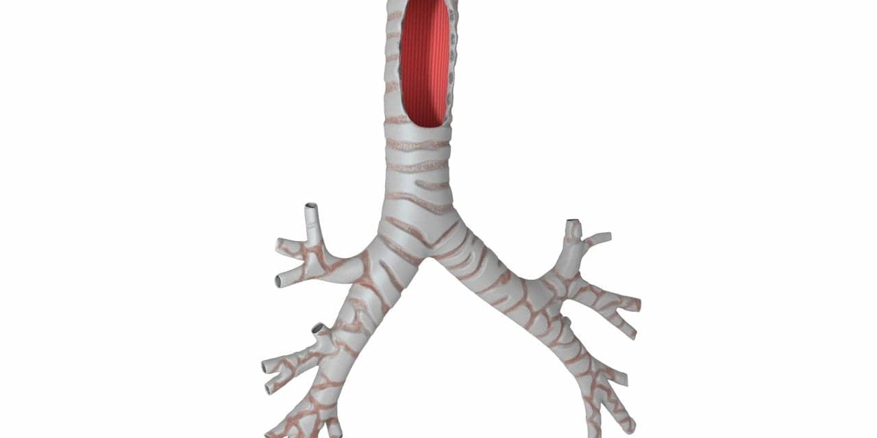
By leveraging computer-aided design and laser-based three-dimensional printing, doctors at the University of Michigan created a customized, bioresorbable tracheal splint using a computed tomographic image of the patient’s airway. Twenty-one days after the procedure, the 20-month-old with tracheobronchomalacia was off ventilator support and has not had breathing trouble since then, according to clinicians. The study appears in the current issue of the New England Journal of Medicine.
“It was amazing,” said Glenn Green, MD, associate professor of pediatric otolaryngology at the University of Michigan. “As soon as the splint was put in, the lungs started going up and down for the first time and we knew he was going to be OK.”

Green and his colleague, Scott Hollister, PhD, professor of biomedical engineering and mechanical engineering and associate professor of surgery at U-M, obtained emergency clearance from the FDA to create and implant a tracheal splint for the child made from a biopolymer called polycaprolactone. The splint was sewn around the patient’s airway to expand the bronchus and give it a skeleton to aid proper growth. Over the course of about three years the splint will be reabsorbed by the body, according to researchers.
“Our vision at the University of Michigan Health System is to create the future of health care through discovery. This collaboration between faculty in our Medical School and College of Engineering is an incredible demonstration of how we achieve that vision, translating research into treatments for our patients,” says Ora Hirsch Pescovitz, MD, executive vice president for medical affairs and CEO of the U-M Health System.
The image-based design and 3D biomaterial printing process can be adapted to build and reconstruct a number of tissue structures, according to Green and Hollister, who have already utilized the process to build and test patient specific ear and nose structures in pre-clinical models. In addition, the method has been used by Hollister with collaborators to rebuild bone structures (spine, craniofacial and long bone) in pre-clinical models.


