Capnography appears to be the most effective monitoring aid for detecting respiratory compromise in patients during mild sedation.
By Kevin Dang and Peter H. Breen, MD, FRCPC
For patients undergoing ambulatory procedures, monitoring of ventilatory status is fundamental. The administration of sedative/analgesic drugs may cause respiratory depression in the ambulatory setting, and the ensuing hypoxia is a common complication.1-4 Early detection of hypoventilation or apnea is critical to the prevention of the complications of hypoxia.5,6
The pulse oximeter is the most commonly used monitor in the ambulatory setting to detect hypoxia. Pulse oximetry measures arterial blood oxygen saturation (Spo2), generally using a digit or ear sensor. Respiration can cease for several minutes, however, before oxygen stores in the lung are consumed (especially during the breathing of supplemental oxygen) and before Spo2 actually begins to decrease.2,6,7,8 In contrast, capnography, the breath-by-breath graphical measurement of the partial pressure of carbon dioxide (Pco2) at the airway opening,9,10 may provide the earliest warning of impending respiratory insufficiency.7,11 An obstructed airway will result in the absence of the expiratory capnogram. Depression of central respiratory drive will result in decreased respiratory rate with or without decreased tidal volume; both cause hypoventilation, increased alveolar Pco2 (Paco2), and increased end-tidal Pco2 (Petco2).8,9 Accordingly, the ability to detect hypercarbia or apnea will alert the clinician to the presence of respiratory compromise, allowing for intervention before the development of hypoxemia.
Capnography
Carbon dioxide is produced by metabolism in body tissues, is transported in blood through venous return to the respiratory system, and is eliminated by cyclical expiration from the lungs.9 Capnography is the measurement, at the airway opening, of airway gas Pco2 as a function of time over each respiratory cycle, with results displayed graphically.
The normal capnogram9,10 is shown in Figure 2, page 28. Phase I (the inspiratory baseline) is inspiration, which normally is devoid of carbon dioxide. The presence of carbon dioxide in inspired gas (carbon dioxide rebreathing) will raise the inspiratory baseline. Phase II (the expiratory upstroke) is the transition between anatomical dead space, which does not participate in gas exchange, and carbon-dioxide–containing alveolar gas from the respiratory bronchioles and alveoli. Phase III (the alveolar plateau) represents most of the exhalation from the alveolar compartment. The alveolar plateau normally has a gentle positive slope. The end of the plateau is the end-tidal Pco2 (Petco2), which represents the Pco2 of the last alveolar gas exhaled at the airway opening. Phase IV (the inspiratory downstroke) is the beginning of the next inspiration.
Capnometers can measure airway carbon dioxide in two ways.9,12 The mainstream (in-line) capnometer incorporates an infrared light source and detector into an airway adapter, which is generally attached in a breathing circuit near a patient face mask, laryngeal mask airway, or endotracheal tube. There is also a disposable self-
contained mainstream capnometer airway adapter that incorporates a pH-sensitive disk (a colorimeter) that changes color semiquantitatively with changes in airway carbon dioxide during the respiratory cycle.13,14 Although the mainstream capnometer has fast measurement response, the airway adapter is delicate; can add significant dead space, bulk, and weight at the airway opening13; and cannot easily monitor the patient who is awake and who has no airway device.
Sidestream capnography provides fast and simple monitoring of carbon dioxide in the spontaneously breathing patient, awake or sedated, and it is almost always used for this purpose. The sidestream capnometer incorporates a low–internal-volume sampling tube connected to or placed at the airway opening. The sidestream capnometer continuously aspirates gas from the patient’s airway device, mouth, or nares into a measuring curette (usually using an infrared measurement technology) that is contained in a separate monitor unit. The time needed to aspirate gas through the sampling tube creates a delay in response time, called the transport delay.15 This delay is increased by lower sampling flow rates; larger sampling tube diameters; longer tube lengths; or partial blockage of the sampling line by secretions, water condensation, or kinking of the tubing. At the same time, axial mixing of gas in the sampling tube can have the effect of averaging the capnogram waveform (decreasing dynamic response),9,15 which can raise the inspiratory baseline. It can also decrease the height of the alveolar plateau and Petco2 and alveolar ventilation is overestimated. Axial mixing of gas is exacerbated in sampling-tube systems with higher internal volumes. Dilution of the sample by surrounding gases such as supplemental oxygen can also decrease the Petco2.7basic pieces of information.9 First, inspection of the waveform shape can detect ventilatory patterns (slow respiratory rate or slow exhalation) and specific abnormalities (blunting of the expiratory upstroke during airway obstruction or an elevated baseline during carbon dioxide rebreathing). Second, Petco2 generally reflects the Paco2 and, hence, the Paco2. Accordingly, in the absence of changes in peripheral tissue carbon dioxide production or changes in the amount of ventilated but unperfused lung (alveolar dead space, which by itself can decrease Petco2),16 a high Petco2 suggests hypoventilation and a low Petco2 suggests hyperventilation.
Monitoring the Capnogram
Several gas-sampling methods using sidestream capnometry have been described; they obtain values for Petco2 that reasonably approximate Paco2. Through modified nasal cannulae, Bowe et al17 insufflated oxygen into one nostril and monitored gas exhaled from the other side, with reliable measurement of Petco2. Other studies18-21 have yielded similar findings using modified nasal cannulae in both adult and pediatric patients. Factors such as mouth breathing, nasal airway obstruction, and administration of oxygen through the ipsilateral naris, however, can result in an underestimation of Petco2 or the false diagnosis of an apneic episode.5,17,18,19,21
To account for both oral and nasal breathing, several monitoring strategies have been developed. First, accurate values for Petco2 can be measured using a nasopharyngeal sampling catheter.5,22 A tube is inserted inferior to the soft palate, thus eliminating the issue of oral versus nasal breathing. The sampling catheter can become blocked with secretions, however. If it is inserted too deeply, the catheter may not be well tolerated in the patient who is awake, and it could induce retching.5 Second, the sampling line can be placed loosely inside an oxygen face mask5,23 in which carbon dioxide sampling accuracy does not depend on the route of breathing. The accuracy of Petco2, however, is inversely related to the supplemental-oxygen flow rate. For example, gross underestimates of Petco2 and poor-quality capnogram waveforms can occur in patients with small tidal volumes and supplemental oxygen flow rates of 6 to 8 L/min.5,24 Nasal/oral sampling systems may provide accurate capnography with improved patient comfort.5,25,26
Clinical Studies
A number of ambulatory clinical studies,5,6,8,20,21,27 including studies of procedures performed under sedation in dental offices and emergency departments, have shown capnography to be an important monitor of the ventilatory state. In studies8,21,27 of pediatric dental patients, it was concluded that the identification of abnormal trends (such as decreasing respiratory rates) on the capnogram can facilitate immediate detection of airway compromise before potential arterial blood desaturation occurs. Croswell et al21 compared three types of monitoring–capnography, pulse oximetry, and clinical observation–in sedated pediatric dental patients. Capnography provided a minimum 15-second warning of a potential arterial blood desaturation event and was the most sensitive method for detecting airway compromise, especially during deeper levels of sedation. Abramo et al25 used oral/nasal capnometry in pediatric patients during active seizures or in the postictal state; they demonstrated that Petco2 monitoring is a useful predictor of hypercarbia and is more sensitive than pulse oximetry in predicting a trend toward respiratory failure.
Studies7,11,20 in adult patients (awake and sedated) have similarly shown that capnography provides the earliest warning of airway obstruction and respiratory compromise. Wright20 observed that increases in Petco2 usually preceded the decrease in blood oxygen saturation. The ability to glance at the capnogram waveform in order to detect changes in respiratory pattern immediately was considered most beneficial.
Limitations of Capnography
The challenge, in the capnographic monitoring of patients who are awake, is to sample expired gases accurately. Several clinical conditions can lead to the recording of erroneous values for Petco2.7,18,27,28 It may be underestimated in association with small tidal volumes, high respiratory rates, large-diameter nasal cannula tips, and high capnometer-sampling flow rates.
Furthermore, any condition that increases alveolar dead space ventilation will decrease Petco2 because Paco2 is diluted by parallel dead space that contains little carbon dioxide.9,16,22 Factors conducive to generation of alveolar dead space include increased alveolar pressure (positive end-expiratory pressure), decreased pulmonary perfusion pressure (low cardiac output or seated position), and physical blockage of a pulmonary arterial branch (venous air embolism).
Conditions in which Petco2 is underestimated can lead to false-positive alerts of ventilatory compromise. In a study21 of sedated pediatric dental patients, the false-alert rate for respiratory compromise was higher for capnography than for clinical monitoring. Conditions that contributed to a false alert included crying, struggling, nasal congestion, and mouth breathing. In another study8 of pediatric dental patients, capnography resulted in excellent sensitivity (100 percent) but less specificity (92 percent) for ventilation problems. The benefit of capnography in providing a more sensitive signal of respiratory compromise, however, far outweighs the inconvenience of false-positive alerts.
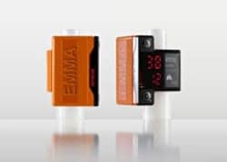
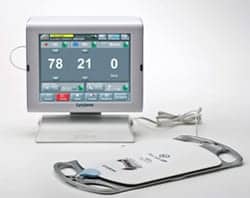
Several portable capnometers are available; these monitors range from lightweight, battery-powered, handheld devices to larger tabletop units. The larger units often require an AC electrical power source, but can be easily carried and moved around an ambulatory clinic. They generally provide a graphic display of the capnogram, along with numerical Petco2 and respiratory-rate data, and some units incorporate pulse oximetry. The price range of these capnography units is $1,000 to $11,000.
Other Clinical Uses
Although it is valuable as a quantitative measure of ventilation, the initial reason that capnography was rapidly accepted into monitoring during anesthesia was its ability to confirm proper placement of the endotracheal tube.9,13 The lung is the only body cavity that receives a continuous input of carbon dioxide from body tissues. After an endotracheal tube placement attempt, cyclical exhaled carbon dioxide during ventilation confirms a patent airway. There are two pitfalls in this application, however.
First, a patent airway does not necessarily mean that the endotracheal tube is correctly positioned (eg, endotracheal tube cuff proximal to the vocal cords). Second, during difficult positive-pressure ventilation with the face mask, the high airway pressure needed may push carbon-dioxide–containing pharyngeal gas into the esophagus and stomach. Then, if the endotracheal tube is mistakenly placed into the esophagus, the ensuing gastric ventilation will result in cyclical–exhaled–carbon dioxide, but with low (generally less than 10 mm Hg) and decreasing values for Pco2. In the ambulatory setting, confirmation of pulmonary ventilation by capnography can be particularly helpful in emergency departments, during cardiopulmonary resuscitation (CPR) on general nursing floors, in the field (although pulmonary blood flow is necessary), and in noisy transport environments.
During cardiac arrest, pulmonary blood flow and carbon dioxide delivery to the lung decrease, with resultant decreases in Petco2, decreases in the elimination of carbon dioxide in exhaled gas, and retention of carbon dioxide in the body.9,16 When native cardiac function and circulation are restored by CPR, the increase in pulmonary blood flow returns a flood of retained carbon dioxide to the lung, and Petco2 markedly increases.29-32 Isserles and Breen16 have shown experimentally that changes in Petco2 correlate directly with changes in pulmonary blood flow. Therefore, portable capnography during CPR in ambulances, in emergency departments, on general floors, and in the field can help confirm the presence of pulmonary blood flow during chest compressions. Furthermore, during constant mechanical ventilation, improved cardiac output achieved by adjusting chest compressions should be signaled by an increase in Petco2. An abrupt increase in Petco2 may signal the return of spontaneous circulation.
Capnography is helpful in the assessment of asthma, where an increased slope in the expiratory plateau provides a rapid, noninvasive, and effort-independent measure of bronchospasm.33,34 The slope of the plateau phase correlates with peak expiratory flow rate, an effort-dependent measurement of airway obstruction. The adequacy of b-agonist bronchodilatory therapy for an asthma exacerbation may be monitored through observation of slope correction in the expiratory plateau.33
Conclusion
Unrecognized respiratory difficulty with resultant hypoxemia is a major contributing factor to morbidity and mortality during conscious sedation in the ambulatory setting. In sedated patients, capnography appears to be the earliest and most effective monitoring aid to the detection of respiratory compromise, and it is recommended in conjunction with pulse oximetry.
RT
Kevin Dang is a visiting senior medical student, Department of Anesthesiology, University of California–Irvine. Peter H. Breen, MD, FRCPC, is associate professor and vice chair, research, in the department. This work was supported by National Institutes of Health grant HL42637. For more information, contact [email protected].
References
1. Bailey PL, Pace NL, Ashburn MA, Moll JWB, East KA, Stanley TH. Frequent hypoxemia and apnea after sedation with midazolam and fentanyl. Anesthesiology. 1990; 73:826-830.
2. Anderson JA, Vann WF. Respiratory monitoring during pediatric sedation: pulse oximetry and capnography. Pediatr Dent. 1988;10:94-101.
3. Charlton JE. Monitoring and supplemental oxygen during endoscopy. BMJ. 1995;310:886-887.
4. Bell GD, McCloy RF, Charlton JE, et al. Recommendations for standards of sedation and patient monitoring during gastrointestinal endoscopy. Gut. 1991;32:823-827.
5. Derrick SJ, Waters H, Kang SW, Cwalina TF, Simmons W. Evaluation of a nasal/oral discriminate sampling system for capnographic respiratory monitoring. Journal of the American Association of Nurse Anesthetists. 1993;5:509-520.
6. Hart LS, Berns SD, Houck CS, Boenning DA. The value of end-tidal CO2 monitoring when comparing three methods of conscious sedation for children undergoing painful procedures in the emergency department. Pediatr Emerg Care. 1997;13:189-193.
7. Kaneko Y. Clinical perspectives on capnography during sedation and general anesthesia in dentistry. Anesthesia Progress. 1995; 42:126-130.
8. Bennett J, Petersen T, Burleson JA. Capnography and ventilatory assessment during ambulatory dentoalveolar surgery. J Oral Maxillofac Surg. 1997;55:921-925.
9. Breen PH. Carbon dioxide kinetics during anesthesia: pathophysiology and monitoring in respiration in anesthesia: pathophysiology and clinical update. Anesthesiology Clinics of North America. Philadelphia: WB Saunders, 1998;16:259-293.
10. Breen PH, Mazumdar B, Skinner SC. Comparison of end-tidal Pco2 and average alveolar expired Pco2 during positive end-expiratory pressure. Anesth Analg. 1996; 82:368-373.
11. Prstojevich SJ, Sabol SR, Goldwasser MS, Johnson C. Utility of capnography in predicting venous carbon dioxide partial pressure in sedated patients during outpatient oral surgery. J Oral Maxillofac Surg. 1992;50:37-39.
12. Pascucci RC, Schena JA, Thompson JE. Comparison of a sidestream and mainstream capnometer in infants. Crit Care Med. 1989;17:560-562.
13. Santos LJ, Varon J, PicAluas L, Combs AH. Practical uses of end tidal carbon dioxide monitoring in the emergency department. J Emerg Med. 1994;12:633-644.
14. Kelly JS, Wilhoit RD, Brown RE, James R. Efficacy of the FEF colorimetric end-tidal carbon dioxide detector in children. Anesth Analg. 1992;75:45-50.
15. Breen PH, Mazumdar B, Skinner SC. Capnometer transport delay: measurement and clinical implications. Anesth Analg. 1994;78:584-586.
16. Isserles SA, Breen PH. Can changes in end-tidal Pco2 measure changes in cardiac output? Anesth Analg. 1991;73:808-814.
17. Bowe EA, Boysen PG, Broome JA, Klein EF. Accurate determination of end-tidal carbon dioxide during administration of oxygen by nasal cannulae. J Clin Monit. 1989;5:105-110.
18. McNulty SE, Roy J, Torjman M, Seltzer JL. Relationship between arterial carbon dioxide and end-tidal carbon dioxide when a nasal sampling port is used. J Clin Monit. 1990;6:93-98.
19. Friesen RH, Alswang M. End-tidal Pco2 monitoring via nasal cannulae in pediatric patients: accuracy and sources of error. J Clin Monit. 1996;12:155-159.
20. Wright SW. Conscious sedation in the emergency department: the value of capnography and pulse oximetry. Ann Emerg Med. 1992;21:551-555.
21. Croswell RJR, Dilley DC, Lucas WJ, Vann WF. A comparison of conventional versus electronic monitoring of sedated pediatric dental patients. Pediatr Dent. 1995;17:332-339.
22. Liu SY, Lee TS, Bongard F. Accuracy of capnography in nonintubated surgical patients. Chest. 1992;102:1512-1515.
23. Inomata S. Early detection of airway obstruction with a capnographic probe attached to an oxygen face mask. Can J Anaesth. 1992;39:744.
24. Ivens D, Verborgh C, Thi HP, Camu F. The quality of breathing and capnography during laryngeal mask and face mask ventilation. Anesthesia. 1995;50:858-862.
25. Abramo TJ, Wiebe RA, Scott S, Goto CS, McIntire DD. Noninvasive capnometry monitoring for respiratory status during pediatric seizures. Crit Care Med. 1997;25:1242-1246.
26. Abramo TJ, Wiebe RA, Scott SM, Primm PA, McIntyre D, Mydler T. Noninvasive capnometry in a pediatric population with respiratory emergencies. Pediatr Emerg Care. 1996;12:252-254.
27. Iwasaki J, Vann WF, Dilley DCH, Anderson JA. An investigation of capnography and pulse oximetry as monitors of pediatric patients sedated for dental treatment. Pediatr Dent. 1989;11:111-117.
28. From RP, Scamman FL. Ventilatory frequency influences accuracy of end-tidal co2 measurements. Analysis of seven capnometers. Anesth Analg. 1988;67:884-886.
29. White RD, Asplin BR. Out-of-hospital quantitative monitoring of end tidal carbon dioxide pressure during CPR. Ann Emerg Med. 1994;23:25-30.
30. Sanders AB. Capnometry in emergency departments. Ann Emerg Med. 1989;18:1287- 1290.
31. Callaham M, Barton C. Prediction of outcome of cardiopulmonary resuscitation from end-tidal carbon dioxide concentration. Crit Care Med. 1990;18:358-362.
32. Garnett AR, Ornato JP, Gonzalez ER, Johnson EB. End-tidal carbon dioxide monitoring during cardiopulmonary resuscitation. JAMA. 1987;257:512-515.
33. Yaron M, Padyk P, Hutsinpiller M, Cairns CB. Utility of the expiratory capnogram in the assessment of bronchospasm. Ann Emerg Med. 1996;28:403-407.
34. You B, Peslin R, Duvivier C, Vu VD, Grilliat JP. Expiratory capnography in asthma: evaluation of various shape indices. Eur Respir J. 1994;7:318-323.