A 40-year-old female with status asthmaticus and respiratory failure was placed on continuous isoflurane ventilation in the ED before safely transported to the medical ICU.
According to the American Lung Association (ALA),1 14.9 million US residents have asthma, and it is the most common chronic illness among children. The Centers for Disease Control and Prevention1 reported a 61% increase in asthma incidence rates between 1982 and 1994. Asthma can be life threatening. According to the ALA,1 more than 5,600 people die in the United States of asthma per year, and mortality increased 45% between 1985 and 1995.
Because increasing numbers of patients were presenting to its emergency departments (EDs) with severe asthma exacerbations, Christiana Care Health System, Newark, Del, created a comprehensive asthma-management program. That program includes the availability of isoflurane for the treatment of patients with severe, life-threatening asthma.
Isoflurane is an anesthetic agent for which use in the treatment of life-threatening asthma has been described.2,4 It causes bronchodilation through a variety of direct and indirect mechanisms, including µ-adrenergic receptor stimulation, direct relaxation of smooth muscle, antagonism to the action of histamine, and interference with hypercapnic bronchoconstriction.3,4 Johnston3 has stated that patients who are already receiving maximum doses of standard bronchodilators may show a response to the addition of isoflurane, and that its rapid action (during the wait for high-dose steroids to take effect) may be lifesaving. At our institution, we have made similar observations about isoflurane therapy for asthma patients.
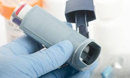
Our isoflurane delivery system includes a ventilator equipped with an isoflurane vaporizer, a vacuum scavenging system and portable tank system, an agent monitor, and an uninterruptible power supply. We designed the system to be used for initiating isoflurane treatment in the ED, with the ability to transport the patient to the medical intensive care unit (ICU) for treatment. A collaborative effort of the respiratory department, anesthesia department, and pulmonary and critical care services formed the basis for the delivery of isoflurane outside the operating room. An RCP team was trained to use the isoflurane system for asthma patients; the anesthesiology department is consulted to oversee patients’ care while they are receiving isoflurane. The respiratory department helped to train the nursing staff in isoflurane therapy, and the isoflurane delivery system is operated and maintained by respiratory care personnel.
Case Summary
A 40-year-old black female presented to the ED with status asthmaticus and respiratory failure. The patient’s history included previous hospital admissions and an intubation for asthma. The patient was spontaneously breathing and in distress, with excessive use of respiratory accessory muscles and severe bilateral inspiratory and expiratory wheezing. On admission, the patient’s pulse rate was 147 beats per minute, her spontaneous respiratory rate was 20 breaths per minute, her oxygen saturation level as measured using pulse oximetry was 97% while she breathed a fraction of inspired oxygen (Fio2) of 100%, and her blood pressure was 179/86. The initial arterial blood gas (ABG) results obtained using a sample taken while she was breathing spontaneously at an Fio2 of 100% were pH, 6.98; Paco2, 122 mm Hg; and Pao2, 87 mm Hg.
Immediately after those results had been determined, the patient was intubated orally using an 8-mm internal diameter endotracheal tube. Medications given on admission were continuous nebulized albuterol sulfate, 10 to15 mg per hour; methylprednisolone, 120 mg every 6 hours; subcutaneous terbutaline, .25 mg; and a lorazepam drip at a rate of .04 mg/kg/hour for sedation. Medications that were added once the patient had been intubated consisted of a cisatracurium drip for paralysis, ketamine, and an increased dosage of lorazepam. The patient could not be ventilated effectively manually due to severe bronchospasm. Initial mechanical-ventilation settings were volume control; tidal volume, 450 mL; set rate, 12 breaths per minute; and Fio2, 100%. Initial ventilating peak pressures were 70 to 80 cm H2O, with a square-wave peak flow set at 33 L per minute. An inspiratory hold maneuver revealed a plateau pressure of 24 cm H2O, which was calculated as equalling an inspiratory resistance of 84 cm H2O/L per second. An expiratory hold maneuver showed an end-expiratory plateau pressure (AutoPEEP) of about 20 cm H2O. End-tidal carbon dioxide values were in the 120 to 129 torr range. The decision was made to ventilate the patient with isoflurane due to the difficulty encountered in ventilating her effectively using conventional means.
Isoflurane treatment was started at 0.5%, with ventilator settings remaining the same. After 15 minutes at 0.5%, the isoflurane was increased to 1% since no side effects such as hypotension or malignant hypothermia were noted. After isoflurane had been in use for 25 minutes, peak pressure had decreased by 10 cm H2O; the Fio2 was reduced to 60%. ABG results indicated a pH of 7.05, a Paco2 of 98 mm Hg, and a Pao2 of 115 mm Hg. As peak pressures decreased another 5 to 10 cm H2O during the next 10 minutes, the tidal volume was increased to 550 mL. The patient was then transported to the medical ICU, using the ventilator with 1% isoflurane and continuous nebulized albuterol during transport. The isoflurane level was increased to 1.5% after transport. Two hours after admission to the ED, the ABG had further improved to a pH of 7.15, a Paco2 of 64 mm Hg, and a Pao2 of 275 mm Hg. Peak pressures, calculated inspiratory resistance, and AutoPEEP (as determined using an expiratory hold maneuver) had all shown dramatic downward trends over the first 12 hours after the initiation of isoflurane; there was also continued improvement in ABG values, as determined using a sample drawn while the patient’s Fio2 was 35% (pH, 7.34; Paco2, 45 mm Hg; and Pao2, 78 mm Hg). The next day the patient was weaned from 1.5% isoflurane in increments of 0.5% over an 8-hour period, with isoflurane being discontinued about 20 hours after it had been initiated. The patient was extubated 44 hours after intubation and was transferred to a general floor bed. The patient was discharged to go home 8 days after admission with no complications.
Asthma is a disease commonly treated in the hospital, and its prevalence is increasing. This disorder can lead to severe illness and death. We have found our isoflurane therapy program for status asthmaticus to be useful in the treatment of these critically ill patients. It has been very helpful to be able to offer isoflurane therapy in the ED and to be able to transport patients safely to the medical ICU with continuous isoflurane ventilation. The RCPs in our department play a key role in the care of these patients during isoflurane therapy.
John Emberger, RRT, is respiratory clinical manager, Christiana Hospital, Newark, Del.
References
1. Trends in Asthma Morbidity and Mortality. Am Lung Assn. November 1998.
2. Parnass SM. Status asthmaticus treated with isoflurane and enflurane. Anesth Analg. 1987;66:193-195.
3. Johnston RG. Isoflurane therapy for status asthmaticus in children and adults. Chest. 1990;97:698-701.
4. Hirshman CA. Mechanism of action of inhalational anesthesia on airways. Anesthesiology. 1982;56:107-111.