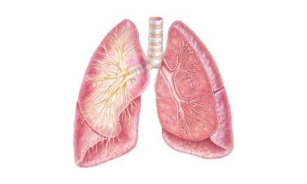
Although COPD and bronchiectasis share many of the hallmark symptoms of obstructive lung diseases, evaluation and treatment for both diseases differ, and diagnosis between the two can be a challenge.
Spirometry has been around for more than 100 years and has served as the primary tool for measuring pulmonary function. While the basic operational principle remains the same, the technology has improved, enhancing performance and interpretation abilities.
However, as advanced as spirometry has become, its ability to differentiate between chronic obstructive pulmonary disease (COPD) and other pulmonary conditions, such as bronchiectasis, has not been fully determined.
Distinct but Related Diagnoses
Although there are some common features between COPD and bronchiectasis that “overlap in circles of the Venn diagram, they are distinct, but related illnesses,” according to Michael W. Sims, MD, COPD program director, assistant professor of clinical medicine at the University of Pennsylvania. He added though that it’s important to make a distinction between the two illnesses to determine the most appropriate course of treatment.
“COPD comprises a group of lung diagnoses that block airflow and make it difficult to breathe. Chronic emphysema and chronic bronchitis are the two most common,” said Sims. “Ninety-five percent of the people who have COPD had been smokers. The disease is characterized by the inability to empty stale air from the lungs. The small airways collapse. There is an accumulation of infection and mucus.” Many patients experience shortness of breath upon exertion, have recurring bronchitis, cough and increased mucus production, although some patients do not present with all symptoms. “The defining feature of COPD is airflow obstruction,” he emphasized.
On the other hand, bronchiectasis can be associated, not just with airflow, but also with damage to the airways. “There might be no airflow obstruction. Its primary characteristic is more anatomical,” he said. “You can see anatomic enlargement on a CT of the chest. If it’s bad enough, you can see it on x-ray.”
Bronchiectasis can be acquired in multiple ways, according to Sims. The disease can be genetic in origin. “Cystic fibrosis is the most common manifestation and leads to thick, tenacious mucus that is not easily cleared,” he said. But in some cases, there is no genetic component, but rather a primary infection, such as tuberculosis or “cousins of tuberculosis,” that cause a “slow, smoldering airway infection.”
Sims further explained that bronchiectasis is a “disease of the immune system,” where the body is not producing enough antibodies, thus triggering immune dysfunction.
“There will be recurring cycles of infection and inflammation in the airway. Infection leads to inflammation, which destroys the airway architecture and enlarges the tubes. This leads to susceptibility of another round of infection. [Bronchiectasis] involves this cycle of infection, inflammation, tissue destruction.”
Sims pointed out that bronchiectasis might also overlap with chronic bronchitis and involve shortness of breath. Clinicians should investigate family history for lung disease or other reasons for recurring cycles of infection.
Determining Diagnosis to Establish Treatment
COPD is definitively linked with cigarette smoking and can be related to environmental smoke exposure. For instance, women who cook over a wood fire in India are exposed to smoke. Those in underserved areas who heat with kerosene run the risk of leaks and can then develop COPD without being a smoker. However, the relationship of smoking in bronchiectasis is less clear. “It’s also not definitively known if environmental factors play a role in bronchiectasis,” Sims said.
What is clear is the diagnostic method, Sims reported. “You can distinguish between the two by evaluating pulmonary function measurements. You can exclude COPD with spirometry, but that’s not true for bronchiectasis,” he said. “You would need an airway imaging CT scan. The best way to distinguish is by clinical criteria.”
Determining a specific diagnosis is important when it comes to identifying the best possible course of treatment. For COPD, the mainstay is inhaled medication that opens the airway and reverses the obstruction, according to Sims. “To some degree, inhaled steroids quiet the inflammatory component and prevent acute exacerbations, which is one of the main factors that lead to disease progression,” he said.
Similarly, clinicians aim to prevent exacerbations in bronchiectasis but with a different pharmacological arsenal. “Inhaled antibiotics suppress bacterial growth and inhaled corticosteroids reduce inflammation,” Sims said. “Both provide maximum clearance of mucus in the airways. And respiratory therapists know of multiple ways to clear the airway using vibration, Acapella, lung flute, mechanical breakup and others.”
Most treatment options for bronchiectasis come out of treatments used for patients with CF. “When you see a new therapy in CF, it will migrate to non-CF bronchiectasis,” Sims said.
Sims pointed out that both COPD and bronchiectasis are irreversible, although it is possible to arrest the progression of the disease. “Bronchiectasis is like an orphan disease. There are specialized centers that treat it,” he said. “COPD is much more common and any medical center can treat it.”
Comorbidity Complicates Conditions
COPD is further complicated by comorbid conditions. Sims said, “COPD is often affected by diseases overlapping by chance.” For instance, a patient could have diabetes and COPD, but there is no defined link between the two diseases. “Some diseases are more commonly associated with COPD and are risk factors for the disease. For example, coronary artery disease is more common in smokers. And any smoker is susceptible to stroke, heart disease and cancers. Lung cancer, in particular, is higher in COPD patients than in the general population.”
Moreover, COPD is associated with muscle weakness and loss of muscle mass, more than would be expected from having lung disease without progressing toward COPD, according to Sims. Depression and anxiety, to a lesser degree, are also common in COPD and can be attributed to permanent progressive loss of breathing function.
Bronchiectasis offers no specific extra pulmonary diagnoses as a global group, Sims reported. “There might be specific things based on what led you to have bronchiectasis. So it could be related to CF and the problems those patients experience,” he said, citing digestive issues and problems with fertility as two common conditions in CF patients.
GOLD Standard
A 2015 update of the Global Initiative for Chronic Obstructive Lung Disease (GOLD) guidelines reported that as the use of CT scan to evaluate patients with COPD is becoming more prevalent, clinicians are identifying “the presence of previously unrecognized radiographic bronchiectasis that appears to be associated with longer exacerbations and increased mortality.”
The guidelines stated, “This ranges from mild tubular bronchiectasis to more severe varicose change, although cystic bronchiectasis is uncommon. Whether this radiological change has the same impact as patients with a primary diagnosis of bronchiectasis remains unknown at present, although it is associated with longer exacerbations and increased mortality.”
More recently, the 2016 GOLD guidelines indicated that when using spirometry clinicians should measure forced vital capacity (FVC) and forced expiratory volume in one second (FEV?), then calculate the ratio of these two measurements.
Theorizing that COPD and bronchiectasis and share some similarities, a 2015 study1 conducted a meta-analysis of all relevant human clinical trials during the first eight months of 2014. They found a mean prevalence of bronchiectasis in COPD patients at 54.3%, with a range of 25.6% to 69%. These two conditions were found more often in males with longer smoking histories. The findings indicate that COPD together with bronchiectasis “should be considered a pathological phenotype of COPD, which may have a predictive value.”
Other researchers have found that bronchiectasis is prevalent in patients with moderate-to-severe COPD.2 This connection was diagnosed after high-resolution CT scan and not by using spirometry.
While CT scan is typically used to diagnose bronchiectasis, spirometry has proven to be a means of detecting exacerbations in patients with the condition. A 2015 study3 evaluated spirometry during three stages: steady state, exacerbations and convalescence. On spirometry, FVC, FEV1 and maximum mid-expiratory flow worsened during exacerbations and recovered during convalescence.
Enhanced Technology
Unlike previous models that relied on a pen drawing a graph on paper as the patient blows into the chamber, today’s spirometers use technology to guide the patient during the testing process, perform the test and interpret the results using ancillary tools. Rich Rosenthal, vice president, US Sales and Marketing, Vitalograph, emphasizes though that the results are “computer suggested and interpretation is not valid without the signature of a licensed medical professional.”
PC-based spirometers, which can be handheld or desktop models, are considered “top of the line,” according to Rosenthal. “You plug the device into a USB port in the computer, do the test using software that displays on the screen in real time,” he said. Newer devices provide motivational tools for the patient. Incentive graphics and animation helps the patient set goals.
Some models package the software in a standalone box that resembles a large laptop and are typically used in a medical workstation. Rosenthal explained that in addition to the spirometer this device integrates a wireless electrocardiograph, blood pressure cuff, pulse oximeter and weight scale.
Other devices, like Vitalograph’s Micro, have a touch screen and removable head. When connected to a computer, the device will produce a PDF report and also store the data in the patient’s electronic medical record. Although the device does not have as much storage capacity as other models, its price tag is appealing to many respiratory therapy departments with tight budgets.
The Food and Drug Administration (FDA) recently approved Vitalograph’s In2itive, a spirometer that sits in a cradle connected to a computer. “The software allows you to print reports or you can send it to the Spirotrac software,” said Rosenthal. The device also has a removable flow head so the patient can view the screen while performing the test.
Regardless of device, Rosenthal noted that the quality of the firmware determines the quality of the test. For example, a device with sophisticated software can detect whether the amount of breath exhaled is sufficient and has reached the six-second mark. It also detects if there is good rise time or if the maneuver was forced or done with hesitation.
Whether spirometry or CT scan is used, the best way to tell which disease a patient has is to consult an expert in the field. “A trained pulmonologist has learned the subtleties and knows how to diagnose and treat the illness,” Sims said. RT
Phyllis Hanlon is a contributing writer to RT. For further information, contact [email protected]
References
-
Ni Y, Shi G, Yu Y et al. “Clinical characteristics of patients with chronic obstructive pulmonary disease with comorbid bronchiectasis: a systemic review and meta-analysis.” International Journal of COPD. 2015:10 1465-1475.
-
Martinez-Garcia MA, de la Rosa Carrillo D, Soler-Cataluña JJ, et al. “Prognostic value of bronchiectasis in patients with moderate-to-severe chronic obstructive pulmonary disease.” Am J Respir Crit Care Med. 2013 Apr 15;187(8):823-31. Doi: 10.1164/rccm.201208-1518OC.
-
Guan WJ, Gao YH, Xu G, et al. “Inflammatory Responses, Spirometry, and Quality of Life in Subjects With Bronchiectasis Exacerbations.” Respir Care. 2015 Aug;60(8):1180-9. doi: 10.4187/respcare.04004. Epub 2015 Jun 9.


