
RTs must be alert to obstructions and malpositioned endotracheal tubes during HFV.
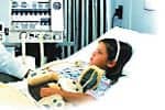 Scenario: An infant’s respiratory support is being provided via high-frequency ventilation (HFV). All seems well, but arterial blood-gas (ABG) analysis indicates a Paco2 of 93 mm Hg. An RT arrives at the patient’s bedside immediately and notes that the infant’s chest is not wiggling (continuous chest vibrations that are equal bilaterally indicate adequate HFV support). The RT ensures that the endotracheal tube’s position is optimal and clears the airway of secretions. The chest wiggle returns, and the patient’s big toes wiggle as well; recovery is rapid.
The same sequence of events might also occur with an adult HFV patient. In this scenario, airway secretions and other tube restrictions had dramatically attenuated the amplitude of HFV support.1,2 Mean airway pressure was sustained, and the Pao2 remained relatively stable. Upon superficial observation, all seemed well because the Pco2 was not being monitored, and the caregiver who drew the ABG sample was not watching for the chest wiggle. As ventilation diminished, chest wiggle stopped and carbon dioxide built up; the RT had seen this happen many times before and quickly remedied the situation. The care team should be cautious about changing Paco2 levels. A rapid reduction of Paco2 back to baseline levels (under permissive hypercapnea, a value in the low 50s) may be detrimental to the infant’s nervous system.3
Another mistake, made in the care of either this infant or any other HFV patient, would have been to respond incorrectly to ABG results. An experienced RT can protect the patient from this problem. If the first response to hypercapnea had been to increase the set amplitude, once the secretion problem had been corrected, the Paco2 could have been driven precipitously downward, past the desired level and into the single digits.
Secretion Control
Use of HFV rather than conventional ventilation may be beneficial in reducing airway inflammation.4 Many aspects of HFV airway maintenance apply to care from birth through adulthood. Conventional ventilation using large tidal volumes provides enough inspiratory time (0.3 seconds to more than 1 second) to penetrate light secretions. Conventional ventilation is usually effective at breathing rates of 10 to 40 breaths per minute (and at more rapid rates for infants). HFV delivers high respiratory rates (420 to 900 breaths per minute), low tidal volumes (similar to dead-space volumes), and short inspiratory times (approximately 0.02 seconds). HFV is more affected than conventional ventilation by secretions and other obstructions. HFV may not provide inspiratory times that are long enough to penetrate secretions sufficiently. Successful HFV depends on optimal transmission of amplitude (pressure change) into the lung. Normally, even with clear airways, most of the HFV amplitude is lost in transmission. Ideally, enough amplitude will reach the lung to provide effective ventilation.
Disconnection from HFV may lead to rapid loss of lung volume (derecruitment).5 RTs at Brigham and Women’s Hospital in Boston try to limit disconnections to once per 12 hours. Auscultation and suction may be done at this time. Collapse, derecruitment, and atelectasis happen within a few breaths at disconnection. Upon reconnection, rerecruitment tends to be much slower; a patient may not recover to baseline oxygen-saturation levels for 10 to 20 minutes. At Brigham and Women’s, we have tried disconnecting the patient and providing re-expanding breaths after derecruitment. Sigh breaths have not been as successful as we had hoped in rerecruiting to the desired baseline functional residual capacity or in improving oxygenation.
We make every attempt to eliminate unnecessary disconnection from the ventilator, and in-line catheters permit the application of suction without disconnection. A study2,5 of rabbits evaluated the differences in mean airway pressure maintenance and oxygenation using a closed suction system versus a disconnected suction process during oscillatory HFV (HFOV). Using a closed suction system maintained mean airway pressure and resulted in less oxygen desaturation. Even in-line suction can cause derecruitment, however. The same suction rules apply as for conventional ventilation. Catheter size should be less than half of the internal diameter of the endotracheal tube. A too-large catheter will draw too much gas from the lung; that will limit ventilation, leading to atelectasis and hemoglobin desaturation. Suction should be applied for a limited time so as to not cause derecruitment. Neonatal in-line devices allow for linear ventilator gas flow from the circuit connection. Mark Rogers, RRT, product manager for high frequency oscillatory ventilation at VIASYS Healthcare (Yorba Linda, Calif), points out that adult in-line devices tend to have a design disadvantage in that their gas flow patterns run at right angles to the ventilator circuit. This angling contributes to attenuation of the ventilator’s amplitude.
Preoxygenation may reduce desaturation during disconnection, suction, or other interruptions of HFOV. Another rabbit study2,3 of the closed suction system demonstrated that lowering the rate of HFOV or increasing amplitude during suction maintained mean airway pressure and oxygenation. There was no evidence of gas trapping with HFOV running. Jean M. Johnson, RRT, clinical sales manager at VIASYS, notes an important concern related to HFV: Erratic lung-pressure fluctuations may be associated with suction during HFV. She suggests that practitioners consider either using alternative manual ventilation or stopping HFV during suction.
Tube Considerations
The size and position of the endotracheal tube may also greatly attenuate HFV amplitude. A change in tube diameter from 2.5 to 3 mm can cause a drop in Paco2 of 10 mm Hg or more. If the diagonal bevel of the tube rides against the back of the trachea, ventilation is attenuated. If the tube slips into a main-stem bronchus, ventilation is diminished. Any bending or twisting of the tube is significant. A misplaced tube is often the cause of a sudden loss of amplitude transmission and chest motion. Newborns have no cuffs to seal their endotracheal tubes to their airways, and an infant who weighs 800 g may, over weeks of ventilatory support, develop a leak around the endotracheal tube. That leak may destabilize HFV amplitude and mean airway pressure. Sometimes leaks set off alarms due to loss of mean airway pressure. Placement of a 3-mm endotracheal tube in an infant weighing less than 1 kg may eliminate the leak.
Some practitioners feel that the infant may not properly qualify for the larger tube until reaching a weight of 1 kg. Adults with tube cuffs have an advantage in controlling seepage of secretions from the pharynx to the trachea. Infants tend to have upper-airway secretions fall into the trachea. For adults, new tubes that have suction control above the cuff may have further advantages in preventing seeping secretions from disrupting HFV’s effectiveness.
Secretions that form a ball-valve obstruction may allow passage of the suction catheter through them, but the HFV impulse will not penetrate them. A quick switch to manual ventilation offers an opportunity to evaluate gas movement through auscultation. A plug may thereby be identified and then removed. Bronchoscopy may also help to identify airway blockages. HFV amplitude, however, is greatly attenuated by the presence of the bronchoscope. It may be necessary to provide conventional ventilation temporarily during the procedure.
Humidification and Jet Techniques
Proper humidification of the airway offers protection from secretion thickening and plugging. It was difficult, with early versions of HFV, to provide humidity. Today, both jet HFV (HFJV) and HFOV humidification systems are adequate. Our experience at Brigham and Women’s is that problems with condensate spilling into the endotracheal tube have not been eliminated by heated wire systems, but merely reduced.
HFJV has been found advantageous in dealing with meconium aspiration.6 According to Evan Richards, clinical specialist for Bunnell Inc (Salt Lake City), HFJV mobilizes meconium and mucus using an action that resembles internal chest physiotherapy; in addition, the sweeping expiratory flow of HFJV appears to facilitate extraction of meconium and mucus. CEO Bert Bunnell has described this mechanism as “the net effect of several HFJV cycles: fresh gas advances down the core airways while exhaled gas moves out along airway walls, which facilitates mucociliary clearance.”6 A special technique is used for suction during HFJV. Jet suction flow is the opposite of HFJV flow. Suction is applied as the catheter is passed into the airway, all the way down to a point just past the tip of the endotracheal tube. Gas flows out of the lungs through the catheter lumen and into the lungs around the walls. This actually prevents machine shutdown and allows fresh gas to flow down the tube during the procedure.
Surfactants, Aerosols, and Monitoring
Some practitioners provide manual ventilation during surfactant delivery; others do not. It is best to complete suction before delivering artificial lung surfactant. This minimizes the interference of secretions, blood, and meconium with surfactant distribution and function. The clinician should also minimize the time that the surfactant-delivery catheter is in the airway, since the catheter can disrupt airway pressure and ventilation. The bolus of surfactant will briefly dampen amplitude at the time of delivery, but as the surfactant is distributed through the lungs, improved compliance will accentuate amplitude, chest wiggle, and ventilation. Suction should not be reapplied until at least an hour has passed after surfactant delivery.
Airway diameter may be improved using aerosolized bronchodilators. It is unfortunate that their delivery is more difficult with HFV than with conventional ventilation. At Brigham and Women’s, we tend to reduce or avoid aerosol delivery to infants receiving HFV. High continuous or bias flow will rush most aerosol past the patient. Placing a metered-dose inhaler’s spacer between the endotracheal tube and ventilator circuit may improve aerosol delivery, but it will also add dead space and decrease ventilation.
A means of monitoring chest vibration is needed. Until one has been developed, continuous ABG monitoring or transcutaneous monitoring will help track carbon dioxide levels.7 The clinician should place the transcutaneous carbon dioxide sensor on the patient before initiating HFV (to determine baseline carbon dioxide levels). Once ventilatory support has been initiated, trends should be followed. Paco2 results can be overly high in circumstances such as poor tissue perfusion. The RT must be good at patient assessment, in addition to providing skilled maintenance and trouble-shooting to ensure accurate data collection.
Conclusion
HFV airway clearance differs from clearance during conventional ventilation. HFOV and HFJV require their own clearance techniques. In the absence of chest wiggle monitoring, RTs must remain alert in detecting obstructions and malpositioned endotracheal tubes.
Michael R. Jackson, RRT-NPS, CPFT, is clinical educator for neonatal and adult respiratory care at Brigham & Women’s Hospital, Boston. Jackson also teaches a course in perinatal and pediatric care at Northeastern University and works as a political advocate in Washington, DC and Massachusetts for the American Association for Respiratory Care.
References
1. Jackson MR, Chuo J. Blood gas and pulmonary graphic monitoring. In: Cloherty JP, Stark AR, eds. Manual of Neonatal Care. Philadelphia: Lippincott Williams & Wilkins; 2004:361-4.
2. Cronin JH. High frequency ventilator therapy for newborns. J Intensive Care Med. 1994;9(2):71-85.
3. Askie LM, Henderson-Smart DJ, Irwig L, Simpson JMl. Oxygen-saturation targets and outcomes in extremely preterm infants. N Engl J Med. 2003;349(10):959-67.
4. Imai Y, Nakagawa S, Ito Y, Kawano T, Slutsky AS, Miyasaka K. Comparison of lung protection strategies using conventional and high-frequency oscillatory ventilation. J Appl Physiol. 2000;91(4):1836-44.
5. Eichenwald E, Stark A. High frequency ventilation: current status. Pediatr Rev. 1999;20(12):e127-33.
6. Bunnell JB. Airway complications of jet ventilation in neonates [comment]. Ann Otol Rhinol Laryngol. 1995;104(9 pt 1):748-9.
7. Courtney SE. Transcutaneous monitoring: back to the future—an important adjunct to care during high frequency ventilation. Neonatology. March 2004. Available at: www.bloodgas.org. Accessed April 4, 2005.


