Miami Valley Hospital performed a mechanical ventilation benchmarking program to determine preweaning and weaning outcomes
Book Review, by Paul Mathews, PhD, RRT, FCCM, FCCP
As a means to continually be cost-effective with quality assured, Miami Valley Hospital (MVH), Dayton, Ohio, chose to embark on a means to not only shorten hospital stays, “Time to Trach,” and time to transfer to an extended care facility (ECF), but also to allow for better quality communication between all allied health care practitioners working on the client’s progress.
As a result, the Hospital Continuum of Care Committee suggested that this area be slated for a benchmarking study. A Mechanical Ventilation Benchmarking Program Committee involving key players from each area of health care involved in mechanical ventilation convened in 1997 and after much planning, education on the program guidelines began in 1998 for all shifts. The study was implemented for 90 days starting on November 30, 1998 to examine the processes for clients requiring mechanical ventilation.
A daily checkoff care flow sheet was designed to document ventilator management, nutrition, activity progression, rest, calm, and comfort. A continuum of care sheet allowed for documentation of procedures (bronchoscopy, tracheostomy placement/plugging/removal, percutaneous endoscopic gastrostomy placement, feeding tube placement, specialty bed, foley insertion/removal, swallowing evaluation, deep venous thrombosis surveillance) and interdisciplinary updates. The nature and scope of the committee was to standardize and improve quality care among all involved with the client’s care.
MVH chose to expand on the work of the National Study Group of the American Association of Critical Care Nurses1 in the areas of mechanical-ventilation preweaning, weaning, and weaning outcomes. Knobel2 described this model; Burns3 described a specific assessment tool called the Burns Weaning Assessment Program. The American Association for Respiratory Care published an entire manual on the topic of weaning protocols.4
A committee convened by the Hospital Continuum of Care Committee examined the processes of care for clients requiring mechanical ventilation. James Murphy, MD, director of the Critical Care/Pulmonary Medicine Department at MVH, chaired the committee. Amy Cline, RRT, director of respiratory services at MVH, served as a member and consultant. This group created the following protocol.
The Benchmarking Mechanical Ventilation Program (BMVP) service begins on the first day of intubation at MVH. Like the Knobel model, the protocol is phase directed, covering preweaning, weaning, and postweaning phases. Its sections include respiratory/airway progression; hemodynamics, including a bronchodilator protocol; nutrition and elimination; activity, rest, and comfort; psychosocial aspects of care, teaching, and discharge planning; continuity of care; and interdisciplinary condition updates.
A 1998-1999 pilot study enabled MVH to analyze tracheostomy demographics. For example, the investigators found that MVH has 100 to 110 tracheostomy patients per year; their mean age is 55 years, and they are released primarily to skilled nursing facilities. The benchmarking study also provided MVH with data on lengths of stay, costs of care, mortality rates, and readmission rates.
The goals set prior to the study were to reduce self-extubations and ventilator-associated pneumonia cases, to reduce the number of ventilator hours, to reduce the number of days that patients spent in bed before mobilization after intubation, to reduce the number of days that elapsed between intubation and physical therapy/occupational therapy consultation, to increase the number of patients receiving uninterrupted sleep, and to decrease the time that elapsed between intubation and tracheostomy.
MVH has achieved success in meeting these goals for several reasons. The majority of MVH clients do not require weaning from mechanical ventilation. MVH is a major trauma center for its area; depending on the cause of intubation, the patient may simply need ventilatory support to be removed in an expeditious fashion, with no gradual weaning necessary. The medical members of the Mechanical Ventilation Benchmarking Program Committee determined that the key factor in successful extubations of this type is recognizing when the patient is ready. This time has usually been reached when the underlying reason for mechanical support has been corrected.
Criteria were set by the committee to help MVH staff to decide whether a given client was ready for weaning. RCPs provide the necessary measurements; if the weaning criteria are met, the patient is ready for a 2-hour trial of continuous positive airway pressure (CPAP) therapy. If CPAP is not tolerated, the patient is returned to use of the pretrial form of ventilatory support.
Weaning consists of retraining the respiratory muscles. The weaning program should be individualized to accommodate the individual’s diagnosis and tolerance. The client is allowed to undergo weaning to point of fatigue (but not to become overly fatigued). Physical therapists know this concept well; the respiratory muscles do not differ from other muscles in their response to fatigue. Specific weaning protocols were designed by the in-house committee.
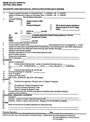
Standard physician-order sheets (Figure 1) provide initial ventilation orders and client parameters. RCPs complete screening to meet weaning criteria by 7 am daily. If the client meets the criteria, the physician is notified that orders are needed. If weaning criteria are not met, physicians can opt for synchronized intermittent mandatory ventilation (SIMV) with pressure support, pressure-support ventilation (PSV), or T-piece/CPAP options.

Preferred weaning hours are 8 am to 9 pm. If PSV and SIMV are used, airway resistance is reevaluated every morning and settings are adjusted accordingly. The mandatory breathing rate is gradually lowered to four breaths per minute. If the client is using positive end-expiratory pressure (PEEP), the change is continued as CPAP on spontaneous respiration. If signs of intolerance are observed, the previous ventilator settings are reinstated (or, if needed, higher settings are used).
With PSV, a tidal volume of 5 to 7 mL/kg should be maintained, along with a respiratory rate that is comfortable for the client. Pressure may be decreased systematically by 3 to 5 cm H2O once per shift if tolerated as long as the client’s spontaneous respiratory rate remains within the safe range. Pressure should be decreased until it equals airway resistance. At this time, the physician can be called for an extubation order. Again, if signs of intolerance begin to show, the client should return to using the previous (or higher) ventilator settings.
If the T-piece/CPAP mode is chosen, the fraction of inspired oxygen (Fio2) will be equal to, or 10% greater than, the baseline level. CPAP will be set to equal the PEEP level. Respiratory vital signs will be assessed 10 to 15 minutes after this change is instituted.

If the client is stable enough to move to a step-down unit (Figure 2, page 56), SIMV with PSV will be used starting the day after the transfer. PSV will be set to equal airway resistance and Fio2 will be decreased to the lowest possible level required by that patient, based on his/her individual needs. PEEP should be set at less than 5 cm H2O. The SIMV rate will be decreased by one breath per minute per day until the client reaches an SIMV rate of four breaths per minute. It will then be changed to both CPAP and PSV, with pressure decreased by 2 to 4 cm H2O each day until it equals airway resistance.
Nonpharmacological intervention should be considered first for pain and sedation when weaning mechanically ventilated clients. Noise control, sleep deprivation, limitation of drugs (benzodiazepines), and lorazepam or morphine may be used for pain and sedation. The physician should be notified if other options do not work; propofol may be useful in trauma cases. A pharmacist is constantly available for consultation with weaning-program staff.
If lorazepam is used, escalating doses should be prescribed. Its onset of action may take place 15 to 20 minutes after its administration, so 1 to 5 mg every 15 minutes for four doses may be required. Its duration of action is 1 to 4 hours, so hyperdynamic clients (such as those with trauma or sepsis) may clear the drug more quickly and elderly patients may clear it more slowly.
If clients require more sedation while receiving continuous infusions of lorazepam, additional bolus doses can be given. The infusion rate of 1 mg per hour may also need to be increased if boluses have been given more frequently than every 1 to 2 hours. The infusion rate should not, however, be increased more than once every 6 hours, so that the maximal effect of the changed infusion rate will be evident. It should be remembered that hypotension can occur if the rate of infusion is too high. Boluses should be infused at a rate no more rapid than 2 mg per minute.
A separate mechanical ventilation flow sheet is provided for use in care documentation by RCPs and nurses. RCPs are to complete screenings for weaning criteria and document their results. The team (consisting of RTs, physicians, nurses, occupational therapists, physical therapists, dieticians, pharmacists, and social workers) determines a goal for the day by evaluating the patient’s weaning candidacy. Goals for pH, Pco2, and pulse-oximetry oxygen saturation (Spo2) should be obtained from the physician’s orders. Baseline Spo2 values may be obtained from previous medical records. The client’s nutrition should be documented, including the rate of feedings or total parenteral nutrition and whether nutritional goals have been met. Use of the aspiration-screening tool should also be included daily and dieticians should monitor for level of consciousness and excessive oral secretions.
The MVH activity progression guideline provides direction for all professionals involved in the client’s care. It consists of five levels; the client should progress to the highest achievable state, from bed rest through head-of-bed elevation through sitting on the side of the bed with the legs dangling and being out of bed in a stretcher chair through bearing his or her own weight in transferring to a canvas wheelchair to fully ambulatory status. The guidelines direct nurses to obtain occupational and physical therapy consultations. Canvas wheelchairs are used in step four because they have more back support and are higher than the chairs found in clients’ rooms. The wheelchairs are to remain available to clients, and occupational or physical therapy staff members who work with the devices are asked to document their use to ensure safety for the next health care worker who comes on the following shift. The use of an individualized activity schedule is encouraged. Continuous sleep lasting at least 4 hours per night is also encouraged, and a nap is recommended during the day. If the client becomes agitated, caregivers should be sure to document the timing and cause of the episode. The continuum of care and interdisciplinary condition update sheet used at MVH provides a weekly summary of the client’s progress toward his or her goals.
Conclusion
After 1 year, the length of stay did not change; however, the time to implement physical therapy, occupational therapy, and feeding regimens did improve greatly upon initial evaluation. “Time to trach” did not change as significantly as the hospital had anticipated due to differences of opinion.
Since the study was targeted to nontrauma patients, the prospective trauma population is now being considered for study to find out if length of stay and costs could be significantly impacted by this population. The number of ventilators run 30 to 35 per day on average and reach as high as 60 in peak months.
The entire MVH respiratory team consists of approximately 150 employees, of whom 110 are involved with adult care and there are 60 employees directly involved with the benchmarking initiative.
Kim Cathcart, MSedA, RN, RRT, is primary care nurse and per diem respiratory care practitioner at Med/Surg Health Center, Miami Valley Hospital, Dayton, Ohio.
References
1. Milliner MP. Ventilator weaning protocols: a comparison study. RT. 2000;13(1):29-32.
2. Knobel A. Weaning from mechanical ventilatory support: a refinement of a model. Am J Crit Care. 1998;7:149-152.
3. Burns S. Burns Weaning Assessment Program (BWAP). Am J Crit Care. 1994;3:12-13.
4. Morris T. Managed care’s impact on academic centers’ research programs. RT. 1999;12:65-68.
| Measurements of Respiratory Mechanics During Mechanical Ventilation
By Paul Mathews, PhD, RRT, FCCM, FCCP Measurements of Respiratory Mechanics During Mechanical Ventilation is a 154-page examination of, as the title states, respiratory mechanics. This volume is packed with useful information and concepts. Two internationally recognized medical scholars wrote the book. The text I received to review appears to be a translation from Italian to English. As such, I expected that there would be some convoluted sentences and paragraphs as Italian words and phrasing may not translate cleanly into English. However, although there were some problems of this sort, these were surprisingly few in number.
The book consists of 10 sections (chapters) each with its own abbreviated reference list. Each chapter is well illustrated with figures illustrating ventilatory waveforms of varying parameters ranging from pressure-volume loops to flow curves. Necessary mathematical equations are presented and discussed at length in each chapter. There is a three-quarter page alphabetic index at the end of the book and almost four pages of table of contents data, which is arranged as a hierarchical outline in a logical, helpful, and comprehensive manner. After a brief preface for each chapter, the authors provide a comprehensive list of abbreviations (2.5 pages) in which many new abbreviations are introduced for later use in the chapter. (This reviewer made a photocopy of the abbreviations and their definitions for easy reference while reading the text—avoiding much page-flipping.) This feature is immediately followed by short discussions of measurement conventions applied during clinical practice in Italy. Further discussion reveals the materials used to develop the data employed in the text. The remainder of the text is divided into three sections: Section A is titled “Basics” inclusive of two chapters; Section B—“Mechanics of the Passive Respiratory System” contains five chapters; and Section C—“Respiratory Mechanics in the Actively Breathing Patient” has three chapters. This is not an easy book to read; both content and layout make it somewhat of a challenge. The need to keep referring back and forth to examine figures discussed in the text and the effort required to decipher the “new” abbreviations slow the reader down and delay comprehension. The level of reading and the depth of discussion are both beyond the ability of most novice readers’ level of comprehension. But for the well-schooled and seasoned practitioner, there is a great deal of valuable information contained in this book. One further problem is the overlaying of “best-fit” lines on the graphs—sometimes they are difficult to discern from the raw data lines. Perhaps in future volumes these best-fit lines could be drawn in another color to help differentiate the source and positions of the lines. Section A (Basics) The authors also introduce many nonstandard abbreviations in this chapter. Examples include V’ instead of the traditional Vdot (for gas flow) and the addition of new nomenclature and symbols. An example would be V’ aw1o for airway opening gas flow. Although these are well defined in the abbreviations list, their unfamiliarity results in a marked slowing of reading speed and comprehension while requiring frequent reference to the abbreviations list in order to understand the discussion. Throughout the book there are repeated references to the plentiful graphics in each chapter. While these added much to the value of the text, references to graphics found on both prior and subsequent pages again required frequent page turning in order to follow the discussion while referencing the appropriate graph. These limitations extend through the remainder of the book; however, the effects become less as one gains familiarity with the nomenclature. In some areas it appears that the authors ignore the differences between the application of classical fluid physics to both compressible and noncompressible fluids. Although these differences are in most cases slight, occasionally, when discussing effects of small volume or pressure changes, they become important. Section B—Mechanics of the Passive Respiratory System Chapter five covers the application of respiratory mechanics by least squares fitting (LSF) in a brief (six pages) and coherent manner. Chapter six, “Respiratory System Time Constants,” is hindered in its presentation by a very convoluted definition of the time constant concept. This is not helped by having graphs 6-1 and 6-2 indicate negative values of V’ aw, while the narrative uses positive or absolute values in its references to the graphic display. Figure 6-3 does not seem to be consistent with the narrative in terms of the intersect of V’ aw = 110 mL/s and Vol = 200 mL. Chapter seven, “Respiratory System Static Pressure-Volume Curves,” does an excellent job of tying the inflection point and best PEEP concepts together in a clearly articulated discussion. This discussion is supported by excellent graphics. Section C—Respiratory Mechanics in the Actively Breathing Patient In chapter eight, figure 8-5 panel 4, this reader could not differentiate the actual from the relaxed curve due to the lines being printed in the same color, type, and size. The discussion of work of breathing was clear and well conceived, as was that of the pressure time product. The authors make a strong case for inclusion of this parameter as a standard measurement. Chapter nine is a short chapter that discusses several of Marini’s methods of determining maximum inspiratory pressure at end inspiration (MIPei) Marini-1 and maximum inspiratory pressure at low lung volume (MIPvl) Marini-2. These are applied to the determination of strength of the inspiratory muscles. Chapter ten discusses the measurement of airway occlusion pressure by two methods, each of which is clearly described. If, as the authors state, the physiologic chain connecting the respiratory center and the respiratory effector organs is assumed to be intact, we can see the effect of neuro stimulus in examination of the P0.1 occlusion pressure. Summary It should also be noted that while the book was sponsored and published by Hamilton Medical AG, few references to ventilators produced by Hamilton are incorporated into the text. Paul Mathews, PhD, RRT, FCCM, FCCP, is associate professor of respiratory care education at the School of Allied Health, University of Kansas Medical Center, Kansas City. |


