Plethysmography plays a key role in assessing functional residual capacity (FRC) in the lung as well as other important pulmonary measures.
By Phyllis Hanlon
Plethysmography has been studied since the mid-1950s, initially to evaluate cardiac output and ventricular activity in animals. In the ensuing years, plethysmography has expanded its focus and plays a key role in assessing functional residual capacity (FRC) in the lung as well as other important measures related to pulmonary condition in humans of all ages.
In 1958, William A. Briscoe and Arthur B. Dubois from the University of Pennsylvania Graduate School of Medicine assessed the airway resistance, airway conductance and lung volume in normal men, women and children, ages 4 to 87.1 Their goal was to establish some baseline measures. They found that age had little effect on the relationship between airway conductance and lung volume; however, they did find a tendency for airway conductance to increase as lung volume decreased.
In 2011, German investigators conducted an analysis and determined that “body plethysmography is a technically demanding, physiologically nontrivial, highly informative, noninvasive method to obtain information on airway obstruction and lung volumes that is not available through spirometry.”2 Moreover, the technique requires just a few minutes to yield “reliable values.”
Fast-forward nearly 60 years and researchers are delving into more sophisticated ways to use plethysmography for patients with compromised pulmonary systems. One approach measures airway resistance (RAW) and specific airway conductance (sGAW) to more accurately arrive at a diagnosis and also differentiate among some pulmonary obstructive illnesses.
Simply put, RAW is “flow resistance of the airways, ie, the ratio of alveolar driving pressure minus mouth pressure to flow rate, computed from sRAW and FRC. RAW indicates the alveolar pressure needed to generate a reference flow rate of one liter per second (1 L/s).”2
sRAW represents the “inverse slope of the plot of flow rate versus box pressure (specific resistance loops) and indicates volume- and resistance-dependent work of breathing needed in order to generate a reference flow rate of 1 L/s.”2
In 2015, a team of Belgium Pulmonary Function Study Investigators3 examined the role RAW and sGAW play in accurate diagnosis of obstructive airway disease and how the two measures factor into differentiating between asthma and chronic obstructive pulmonary disease (COPD). Initially, 976 subjects were enrolled and underwent complete pulmonary function testing and physician examination. Subsequently, 651 subjects were determined to have a final diagnosis of obstructive diagnosis (including COPD, asthma, upper airway obstruction, chronic bronchitis, bronchiolitis, small airway disease, bronchiectasis and cystic fibrosis); 168 were found to have another, non-obstructive respiratory illness; and 157 subjects with no respiratory condition served as healthy controls.
The study intended to “explore the variability of resistance parameters between established diagnoses of respiratory diseases in a general patient sample, and investigate whether measures of resistance can differentiate between the obstructive diseases and further refine the diagnosis of asthma or COPD.”
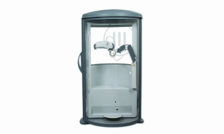
In an effort to achieve a comprehensive evaluation, the investigators used a Sensormedics Whole Body Plethysmograph and Masterscreen Jaeger, both by CareFusion, in addition to a pulmonary function testing station.
At the conclusion of the study, results showed that 47% of all subjects with obstructive airways disease had increased RAW. When less stringent cutoff parameters were used, 37% showed increased resistance. Between 90 and 92% of subjects, depending on cutoff parameters, had low sGAW, which is accurately predictive for obstructive disease.
In spite of promising results, the American Association for Respiratory Care (AARC) has issued guidelines regarding possible limitations when using body plethysmography and urges caution in choosing and applying reference values when validating results.
RT
Phyllis Hanlon is a contributing writer to RT. For further information, contact [email protected].
References
- Briscoe WA, Dubois AB. “The relationship between airway resistance, airway conductance and lung volume in subjects of different age and body size.” Journal of Clinical Investigation. September 1958. 37(9):1279-85. DOI: 10.1172/JCI103715
- Criée CP, Sorichter S, Smith HJ et al. “Body plethysmography – its principles and clinical use.” Respiratory Medicine. 2011. 105, 959-97. Doi:10.1016/j.rmed.2011.02.006.
- Topalovic M, Derom E, Osadnik CR et al. “Airways resistance and specific conductance for the diagnosis of obstructive airways diseases.” Respiratory Research. 2015; 16:88. DOI 10.1186/s12931-015-0252-0.
- http://www.rcjournal.com/cpgs/bplthcpg-update.html